The [KO Validated] SAV1 Antibody (CAB18667) is a high-quality antibody developed for reliable detection and analysis of target proteins. This pathway plays a critical role in controlling organ size, tissue growth, and tumorigenesis, making SAV1 an important target for cancer research.Raised in rabbits, this antibody is highly reactive with human samples and has been rigorously validated for use in Western blot applications. By specifically binding to the SAV1 protein, researchers can easily detect and analyze its expression in various cell types.
This antibody is validated for use in WB, IHC-P, ELISA applications and has demonstrated reactivity against Human, Mouse, Rat samples.
Product Name:
[KO Validated] SAV1 Antibody
SKU:
CAB18667
Size:
20μL, 100μL
Reactivity:
Human, Mouse, Rat
Immunogen:
Recombinant protein (or fragment).This information is considered to be commercially sensitive.
Recommended starting concentration is 1 μg/mL. Please optimize the concentration based on your specific assay requirements.
Synonyms:
SAV, WW45, WWP4, V1
Positive Sample:
HeLa(KO)
Cellular Localization:
Cytoplasm, Nucleus.
Calculated MW:
45kDa
Observed MW:
45kDa
WW domain-containing proteins are found in all eukaryotes and play an important role in the regulation of a wide variety of cellular functions such as protein degradation, transcription, and RNA splicing. This gene encodes a protein with two WW domains, a SARAH domain, and a coiled-coil region and is ubiquitously expressed in adult tissues. This protein binds to MST1 (mammalian sterile 20-like kinase 1) and promotes MST1-induced apoptosis. It has also been shown to bind to HAX1 (hematopoietic cell-specific protein 1 (HS1)-associated protein X-1) and to attenuate the anti-apoptotic effects of HAX1. Studies in human and mouse suggest this gene acts as a tumor suppressor.
Purification Method
Affinity purification
Gene ID
60485
RRID
AB_2862404
Buffer Information
Store at -20℃. Avoid freeze / thaw cycles. Buffer: PBS containing 50% glycerol, preserved with proclin300 or sodium azide, pH 7.3.
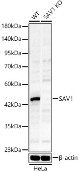
Western blot analysis of lysates from wild type (WT) and SAV1 knockout (KO) HeLa cells, using [KO Validated] SAV1 Rabbit pAb (CAB18667) at 1:1000 dilution. Secondary antibody: HRP-conjugated Goat anti-Rabbit IgG (H+L) (CABS014) at 1:10000 dilution. Lysates/proteins: 25μg per lane. Blocking buffer: 3% nonfat dry milk in TBST. Detection: ECL Basic Kit (AbGn00020). Exposure time: 90s.
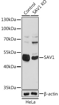
Western blot analysis of lysates from wild type (WT) and SAV1 knockout (KO) HeLa(KO) cells, using SAV1 Rabbit pAb (CAB18667) at 1:1000 dilution. Secondary antibody: HRP-conjugated Goat anti-Rabbit IgG (H+L) (CABS014) at 1:10000 dilution. Lysates/proteins: 25ug per lane. Blocking buffer: 3% nonfat dry milk in TBST. Detection: ECL Basic Kit (AbGn00020). Exposure time: 50s.
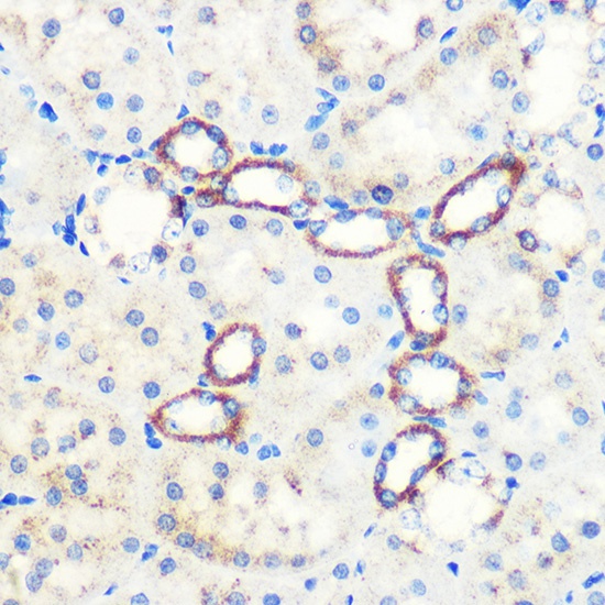
Immunohistochemistry analysis of paraffin-embedded Rat kidney using [KO Validated] SAV1 Rabbit pAb (CAB18667) at dilution of 1:100 (40x lens). Microwave antigen retrieval performed with 0.01M PBS Buffer (pH 7.2) prior to IHC staining.
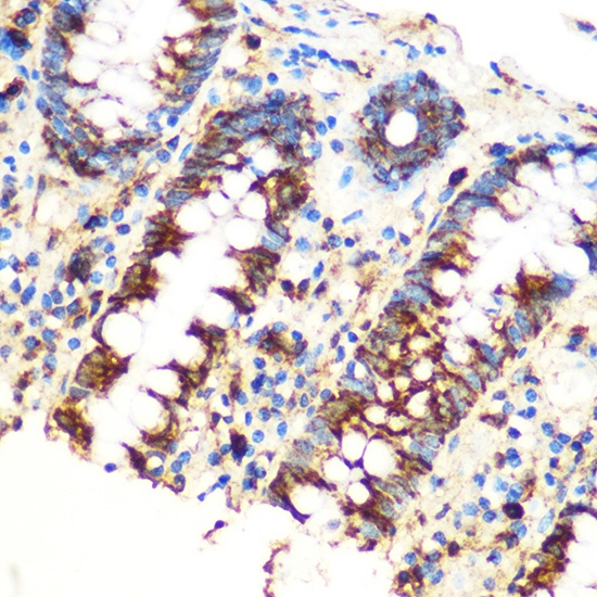
Immunohistochemistry analysis of paraffin-embedded Human colon using [KO Validated] SAV1 Rabbit pAb (CAB18667) at dilution of 1:100 (40x lens). Microwave antigen retrieval performed with 0.01M PBS Buffer (pH 7.2) prior to IHC staining.