The 14-3-3 sigma Antibody (CAB1026) is a high-quality antibody developed for reliable detection and analysis of target proteins. This antibody, produced in rabbits, exhibits high reactivity with human samples and is validated for use in Western blot applications. By specifically binding to the SFN protein, researchers can accurately detect and analyze SFN levels in various cell types, making it an ideal choice for studies in cancer biology and cellular signaling pathways.
This antibody is validated for use in WB, IHC-P, ELISA applications and has demonstrated reactivity against Human, Mouse, Rat samples.
Product Name:
14-3-3 sigma Antibody
SKU:
CAB1026
Size:
20μL, 100μL
Reactivity:
Human, Mouse, Rat
Conjugate:
Unconjugated
Immunogen:
Synthetic peptide. This information is considered to be commercially sensitive.
Recommended starting concentration is 1 μg/mL. Please optimize the concentration based on your specific assay requirements.
Synonyms:
YWHAS, 14-3-3 sigma
Positive Sample:
HeLa, A-549, MCF7, A-431, Mouse lung, Mouse kidney, Rat lung
Cellular Localization:
Cytoplasm, Nucleus, Secreted.
Calculated MW:
28kDa
Observed MW:
28kDa
This gene encodes a cell cycle checkpoint protein. The encoded protein binds to translation and initiation factors and functions as a regulator of mitotic translation. In response to DNA damage this protein plays a role in preventing DNA errors during mitosis.
Purification Method
Affinity purification
Gene ID
2810
RRID
AB_2757787
Buffer Information
Store at -20℃. Avoid freeze / thaw cycles. Buffer: PBS containing 50% glycerol, preserved with proclin300 or sodium azide, pH 7.3.
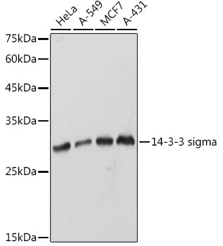
Western blot analysis of various lysates using 14-3-3 sigma Rabbit pAb (CAB1026) at 1:1000 dilution. Secondary antibody: HRP-conjugated Goat anti-Rabbit IgG (H+L) (CABS014) at 1:10000 dilution. Lysates/proteins: 25μg per lane. Blocking buffer: 3% nonfat dry milk in TBST. Detection: ECL Basic Kit (AbGn00020). Exposure time: 1s.
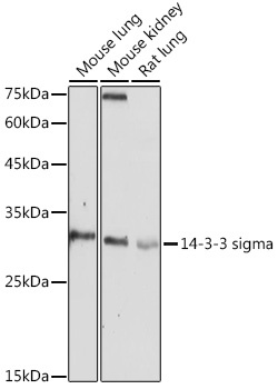
Western blot analysis of various lysates using 14-3-3 sigma Rabbit pAb (CAB1026) at 1:1000 dilution. Secondary antibody: HRP-conjugated Goat anti-Rabbit IgG (H+L) (CABS014) at 1:10000 dilution. Lysates/proteins: 25μg per lane. Blocking buffer: 3% nonfat dry milk in TBST. Detection: ECL Basic Kit (AbGn00020). Exposure time: 180s.
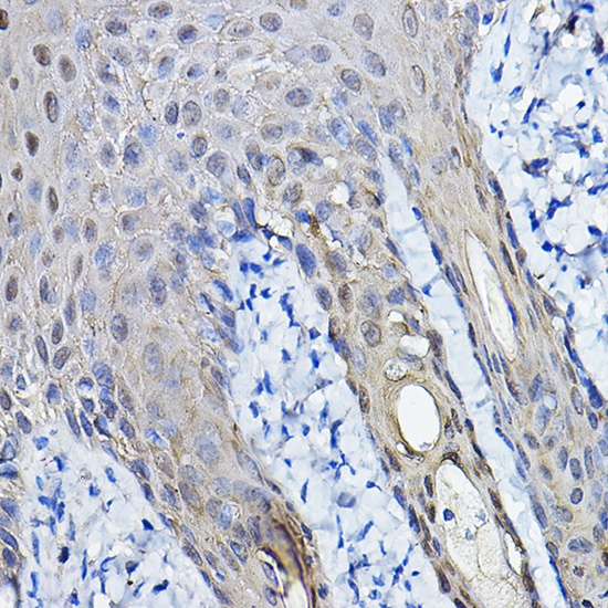
Immunohistochemistry analysis of paraffin-embedded Human skin using 14-3-3 sigma Rabbit pAb (CAB1026) at dilution of 1:100 (40x lens). High pressure antigen retrieval performed with 0.01M Citrate buffer (pH 6.0) prior to IHC staining.
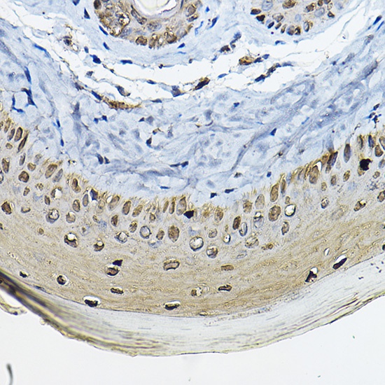
Immunohistochemistry analysis of paraffin-embedded Mouse skin using 14-3-3 sigma Rabbit pAb (CAB1026) at dilution of 1:100 (40x lens). High pressure antigen retrieval performed with 0.01M Citrate buffer (pH 6.0) prior to IHC staining.