The Ku70 Monoclonal Antibody (CAB11223) is a high-quality antibody developed for reliable detection and analysis of target proteins. This antibody is produced in rabbits and is highly specific to human samples, making it ideal for use in various research applications, including Western blotting and immunofluorescence.The KU70 protein is a key component of the DNA-dependent protein kinase complex, which is involved in the repair of DNA double-strand breaks. Dysregulation of KU70 has been linked to genetic instability and the development of various diseases, including cancer.
This antibody is validated for use in WB, IHC-P, IF/ICC, ELISA applications and has demonstrated reactivity against Human, Mouse, Rat samples.
Product Name:
Ku70 Monoclonal Antibody
SKU:
CAB11223
Size:
20μL, 100μL
Reactivity:
Human, Mouse, Rat
Clone Number:
ARC0551
Conjugate:
Unconjugated
Immunogen:
Synthetic peptide. This information is considered to be commercially sensitive.
Recommended starting concentration is 1 μg/mL. Please optimize the concentration based on your specific assay requirements.
Synonyms:
ML8, KU70, TLAA, CTC75, CTCBF, G22P1, Ku70
Positive Sample:
A-549
Cellular Localization:
Chromosome, Nucleus.
Calculated MW:
70kDa
Observed MW:
70kDa
The p70/p80 autoantigen is a nuclear complex consisting of two subunits with molecular masses of approximately 70 and 80 kDa. The complex functions as a single-stranded DNA-dependent ATP-dependent helicase. The complex may be involved in the repair of nonhomologous DNA ends such as that required for double-strand break repair, transposition, and V(D)J recombination. High levels of autoantibodies to p70 and p80 have been found in some patients with systemic lupus erythematosus.
Purification Method
Affinity purification
Gene ID
2547
RRID
AB_2861526
Buffer Information
Store at -20℃. Avoid freeze / thaw cycles. Buffer: PBS containing 50% glycerol and 0.05% BSA, preserved with proclin300 or sodium azide, pH 7.3.
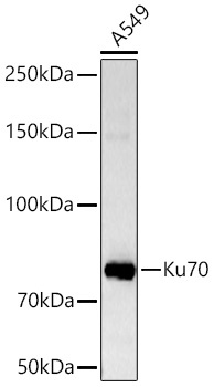
Western blot analysis of lysates from A549 cells using Ku70 Rabbit mAb (CAB11223) at 1:6000 dilution incubated overnight at 4℃. Secondary antibody: HRP-conjugated Goat anti-Rabbit IgG (H+L) (CABS014) at 1:10000 dilution. Lysates/proteins: 25 μg per lane. Blocking buffer: 3% nonfat dry milk in TBST. Detection: ECL Basic Kit (AbGn00020). Exposure time: 10s.
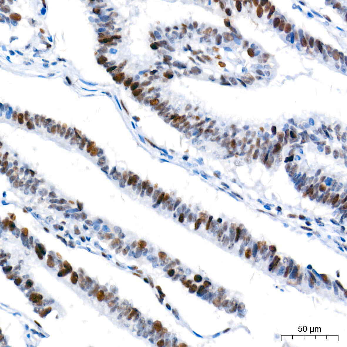
Immunohistochemistry analysis of paraffin-embedded Human colon carcinoma tissue using Ku70 Rabbit mAb (CAB11223) at a dilution of 1:500 (40x lens). High pressure antigen retrieval performed with 0.01M Tris-EDTA Buffer (pH 9.0) prior to IHC staining.
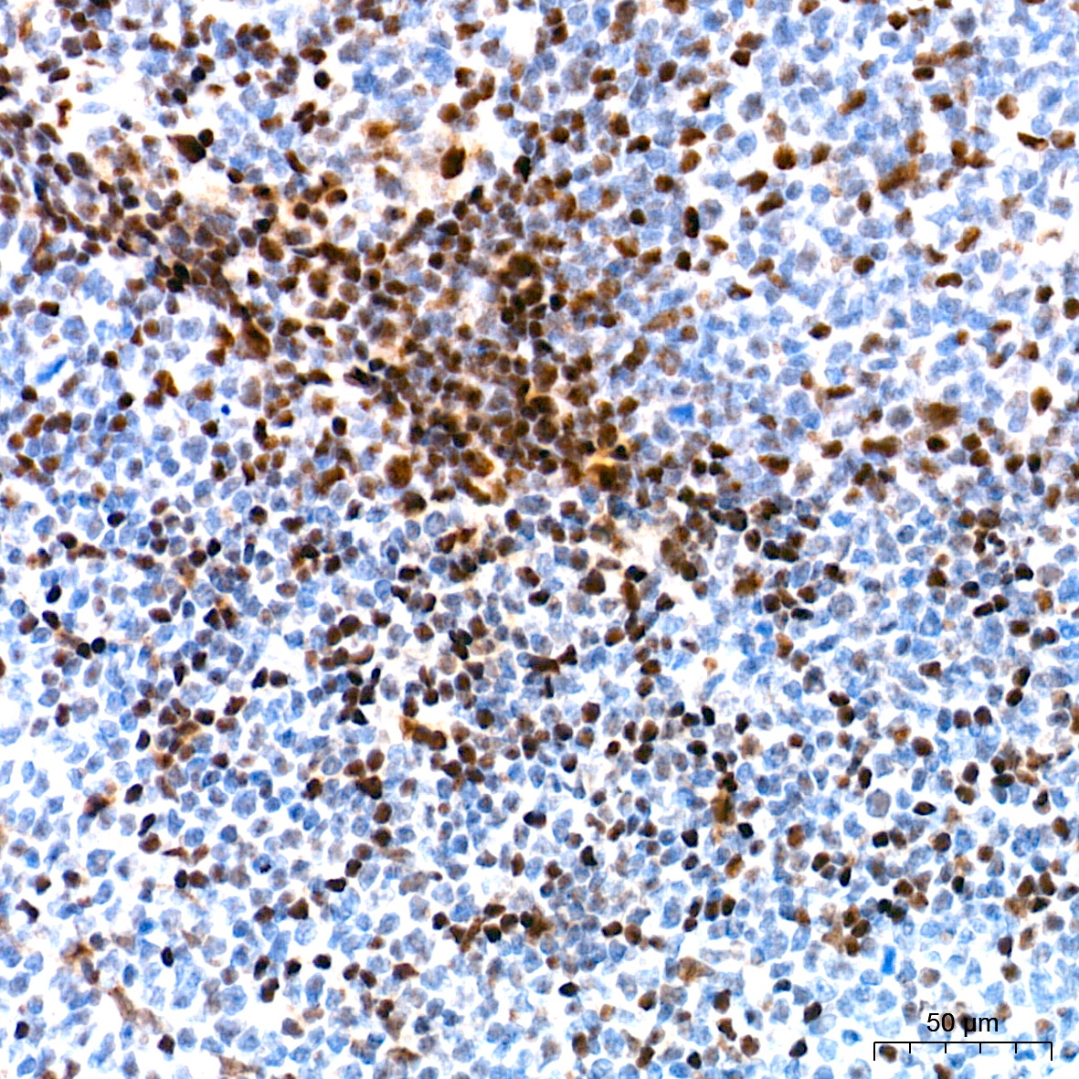
Immunohistochemistry analysis of paraffin-embedded Human tonsil tissue using Ku70 Rabbit mAb (CAB11223) at a dilution of 1:500 (40x lens). High pressure antigen retrieval performed with 0.01M Tris-EDTA Buffer (pH 9.0) prior to IHC staining.
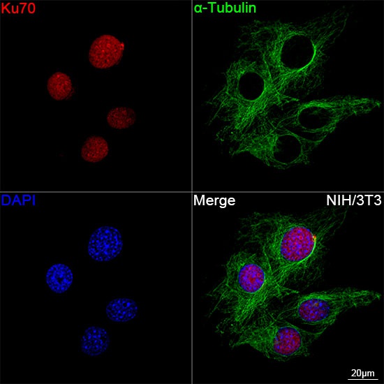
Confocal imaging of NIH/3T3 cells using Ku70 Rabbit mAb (CAB11223, dilution 1:100) followed by a further incubation with Cy3 Goat Anti-Rabbit IgG (H+L) (CABS007, dilution 1:500) (Red). The cells were counterstained with α-Tubulin Mouse mAb (AC012, dilution 1:400) followed by incubation with ABflo® 488-conjugated Goat Anti-Mouse IgG (H+L) Ab (CABS076, dilution 1:500) (Green). DAPI was used for nuclear staining (Blue). Objective: 100x.
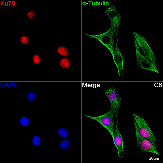
Confocal imaging of C6 cells using Ku70 Rabbit mAb (CAB11223, dilution 1:100) followed by a further incubation with Cy3 Goat Anti-Rabbit IgG (H+L) (CABS007, dilution 1:500) (Red). The cells were counterstained with α-Tubulin Mouse mAb (AC012, dilution 1:400) followed by incubation with ABflo® 488-conjugated Goat Anti-Mouse IgG (H+L) Ab (CABS076, dilution 1:500) (Green). DAPI was used for nuclear staining (Blue). Objective: 100x.