The Ku80 Polyclonal Antibody (CAB24867) is a high-quality antibody developed for reliable detection and analysis of target proteins. This antibody, generated in rabbits, demonstrates high reactivity with human samples and is validated for use in Western blot applications. By binding specifically to Ku80, researchers can easily detect and analyze this important protein in a variety of cell types, making it ideal for studies in genetics, cancer research, and molecular biology.Ku80 is a critical component of the DNA-dependent protein kinase complex that plays a crucial role in repairing DNA double-strand breaks.
This antibody is validated for use in WB, IHC-P, IF/ICC, ELISA applications and has demonstrated reactivity against Human, Mouse samples.
Product Name:
Ku80 Polyclonal Antibody
SKU:
CAB24867
Size:
20μL, 100μL
Reactivity:
Human, Mouse
Conjugate:
Unconjugated
Immunogen:
Recombinant protein (or fragment).This information is considered to be commercially sensitive.
Recommended starting concentration is 1 μg/mL. Please optimize the concentration based on your specific assay requirements.
Synonyms:
Ku80, Ku86, CTC85, CTCBF
Positive Sample:
NIH/3T3, Neuro-2a, HeLa
Cellular Localization:
Nucleus, Nucleus, Nucleolus.
Calculated MW:
83kDa
Observed MW:
83kDa
Predicted to enable several functions, including nucleic acid binding activity; protein C-terminus binding activity; and ubiquitin protein ligase binding activity. Predicted to contribute to 5'-deoxyribose-5-phosphate lyase activity and double-stranded telomeric DNA binding activity. Acts upstream of or within several processes, including cellular response to leukemia inhibitory factor; hematopoietic stem cell differentiation; and positive regulation of neurogenesis. Located in cytoplasm and nucleus. Is expressed in thymus primordium. Human ortholog(s) of this gene implicated in chronic obstructive pulmonary disease and multiple myeloma. Orthologous to human XRCC5 (X-ray repair cross complementing 5).
Purification Method
Affinity purification
Gene ID
22596
Buffer Information
Store at -20℃. Avoid freeze / thaw cycles. Buffer: PBS containing 50% glycerol, preserved with proclin300 or sodium azide, pH 7.3.
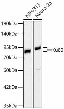
Western blot analysis of various lysates using Ku80 Rabbit pAb (CAB24867) at 1:900 dilution. Secondary antibody: HRP-conjugated Goat anti-Rabbit IgG (H+L) (CABS014) at 1:10000 dilution. Lysates / proteins: 25 μg per lane. Blocking buffer: 3 % nonfat dry milk in TBST. Detection: ECL Basic Kit (AbGn00020). Exposure time: 90s.
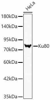
Western blot analysis of lysates from HeLa cells using Ku80 Rabbit pAb (CAB24867) at 1:900 dilution. Secondary antibody: HRP-conjugated Goat anti-Rabbit IgG (H+L) (CABS014) at 1:10000 dilution. Lysates/proteins: 25 μg per lane. Blocking buffer: 3% nonfat dry milk in TBST. Detection: ECL Basic Kit (AbGn00020). Exposure time: 1s.
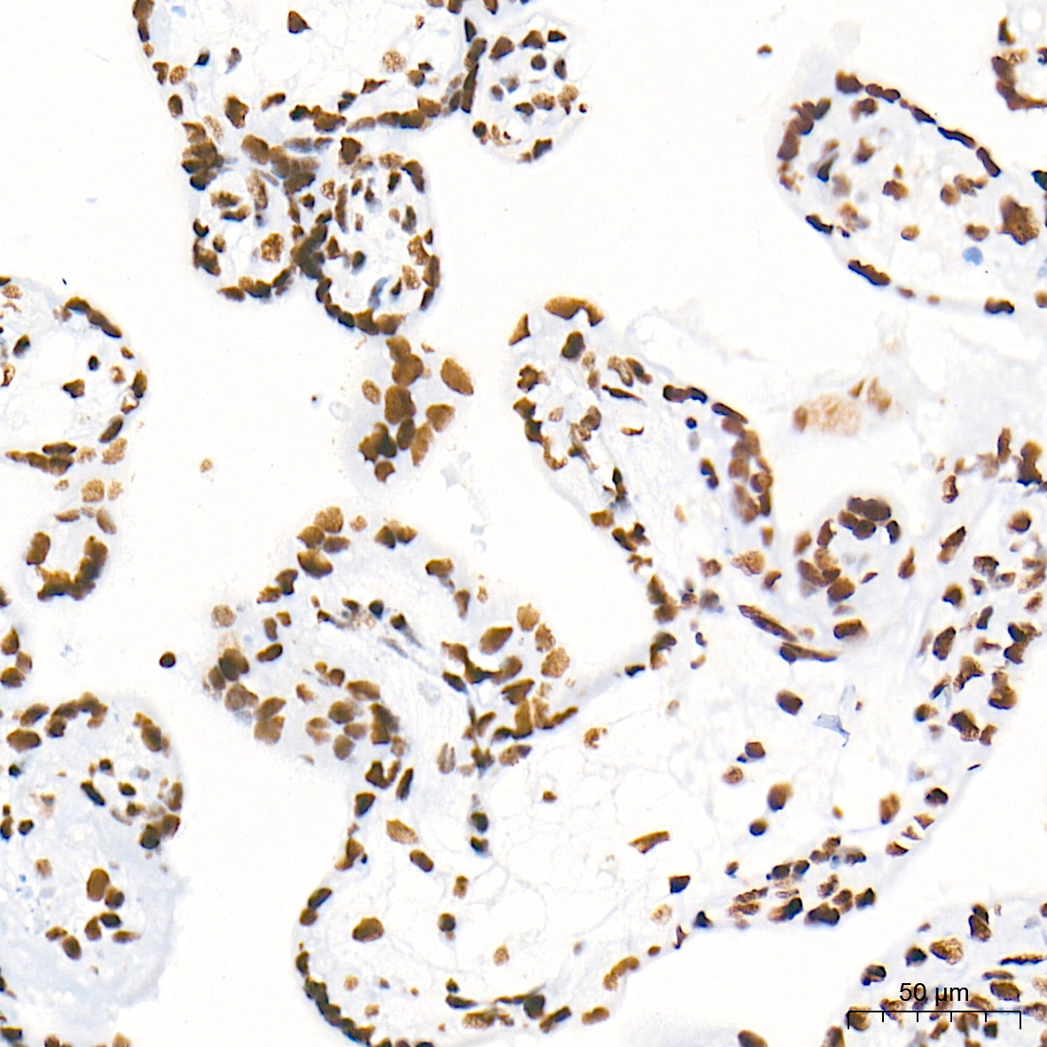
Immunohistochemistry analysis of paraffin-embedded Human placenta tissue using Ku80 Rabbit pAb (CAB24867) at a dilution of 1:4000 (40x lens). High pressure antigen retrieval performed with 0.01M Tris-EDTA Buffer (pH 9.0) prior to IHC staining.
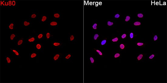
Immunofluorescence analysis of HeLa cells using Ku80 Rabbit pAb (CAB24867) at a dilution of 1:100 (40x lens). Secondary antibody: Cy3-conjugated Goat anti-Rabbit IgG (H+L)(CABS007) at 1:500 dilution. Blue: DAPI for nuclear staining.
Amisten et al.
The DNA repair factor ku80 binds and activates the adhesion receptor ELTD1/ADGRL4
")
")


