lacZ Monoclonal Antibody (MACO0616) is a premium monoclonal that offers outstanding performance and reliability for demanding research applications. Rigorously validated for ELISA, WB, IF, IP, FC, this antibody ensures consistent, reproducible results across multiple experimental platforms. Demonstrates excellent reactivity with Escherichia coli samples, providing researchers with confidence in cross-species compatibility. Conveniently packaged in 50ul format to meet your experimental needs. For optimal performance, store at -20°C or -80°C and maintains stability for 12 months. Backed by rigorous quality control testing to ensure superior performance in your critical research applications.
Product Name:
lacZ Monoclonal Antibody (MACO0616)
SKU:
MACO0616
Size:
50μl
Isotype:
IgG1
Host Species:
Mouse
Reactivity:
Escherichia coli
Immunogen:
Recombinant Escherichia coli Beta-galactosidase protein (2-1024AA)
Immunogen Species:
Escherichia coli
Uniprot No:
P00722
Form:
Liquid
Tested Applications:
ELISAWBIFIPFC
Recommended Dilution:
WB 1:1000-1:64000, IF 1:50-1:200, IP 1ul-4ul, FC 1:100-1:300
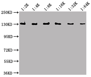
Western Blot Positive WB detected in Recombinant protein All lanes: lacZ antibody at 1:2000, 1:4000, 1:8000, 1:16000, 1:32000, 1:64000 Secondary Goat polyclonal to Mouse IgG at 1/50000 dilution Predicted band size: 117 kDa Observed band size: 130 kDa
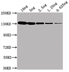
Western Blot Positive WB detected in Recombinant protein (10ng, 5ng, 2.5ng, 1.25ng, 0.625ng) All lanes: lacZ antibody at 1:2000 Secondary Goat polyclonal to Mouse IgG at 1/50000 dilution Predicted band size: 117 kDa Observed band size: 130 kDa
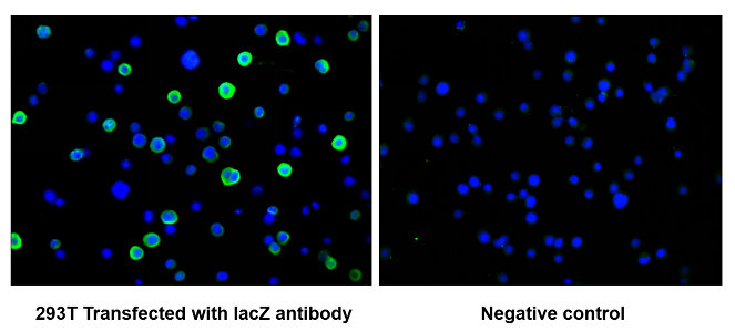
Immunofluorescence staining of 293T cells transfected with MACO0616 at 1:100, counter-stained with DAPI. The cells were fixed in 4% formaldehyde and blocked in 10% normal Goat Serum. The cells were then incubated with the antibody overnight at 4°C. The secondary antibody was Alexa Fluor 488-congugated AffiniPure Goat Anti-Rabbit IgG(H+L). The image on the right is the 293T cells transfected without lacZ antibody
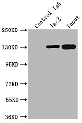
Immunoprecipitating lacZ in 293T transfected whole cell lysate Lane 1: Mouse control IgG instead of MACO0616 in 293T transfected whole cell lysate Lane 2: MACO0616 (2µl) + 293T transfected whole cell lysate (500µg) Lane 3: 293T transfected whole cell lysate (20µg) For western blotting, the blot was detected with CSB-MA000021M0m at 1:2000, and a HRP-conjugated Protein G antibody was used as the secondary antibody at 1:2000
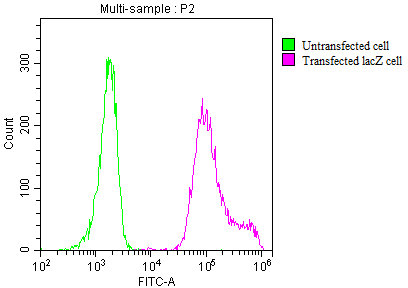
Overlay histogram showing 293T transfected cells (red line) or untransfected cells (green line) stained with MACO0616 at 1:200. The cells were incubated in 1x PBS /10% normal goat serum to block non-specific protein-protein interactions followed by primary antibody for 1 h at 4°C. The secondary antibody used was FITC goat anti-mouse IgG(H+L) at 1/200 dilution for 1 h at 4°C.


 ELISA Kit (HUFI03337)")


