The Lamin B2 Monoclonal Antibody (CAB5001) is a high-quality antibody developed for reliable detection and analysis of target proteins. This antibody, produced in rabbits, demonstrates high specificity and sensitivity for detecting Lamin B2 in human samples, making it a reliable tool for various applications, including Western blot analysis.Lamin B2 is a crucial component of the nuclear lamina and plays a vital role in maintaining nuclear stability and integrity. Dysregulation of Lamin B2 has been implicated in various diseases, including cancer and genetic disorders.
This antibody is validated for use in WB, IHC-P, IF/ICC, ELISA applications and has demonstrated reactivity against Human, Mouse, Rat samples.
Product Name:
Lamin B2 Monoclonal Antibody
SKU:
CAB5001
Size:
20μL, 100μL
Reactivity:
Human, Mouse, Rat
Clone Number:
ARC1252
Conjugate:
Unconjugated
Immunogen:
Synthetic peptide. This information is considered to be commercially sensitive.
This gene encodes a B type nuclear lamin. The nuclear lamina consists of a two-dimensional matrix of proteins located next to the inner nuclear membrane. The lamin family of proteins make up the matrix and are highly conserved in evolution. During mitosis, the lamina matrix is reversibly disassembled as the lamin proteins are phosphorylated. Lamin proteins are thought to be involved in nuclear stability, chromatin structure and gene expression. Vertebrate lamins consist of two types, A and B. Mutations in this gene are associated with acquired partial lipodystrophy.
Purification Method
Affinity purification
Gene ID
84823
RRID
AB_2863410
Buffer Information
Store at -20℃. Avoid freeze / thaw cycles. Buffer: PBS containing 50% glycerol and 0.05% BSA, preserved with proclin300 or sodium azide, pH 7.3.
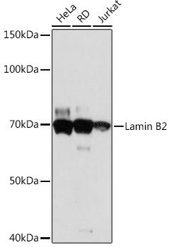
Western blot analysis of various lysates using Lamin B2 Rabbit mAb (CAB5001) at 1:1000 dilution. Secondary antibody: HRP-conjugated Goat anti-Rabbit IgG (H+L) (CABS014) at 1:10000 dilution. Lysates/proteins: 25μg per lane. Blocking buffer: 3% nonfat dry milk in TBST. Detection: ECL Basic Kit (AbGn00020). Exposure time: 30s.
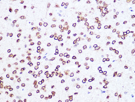
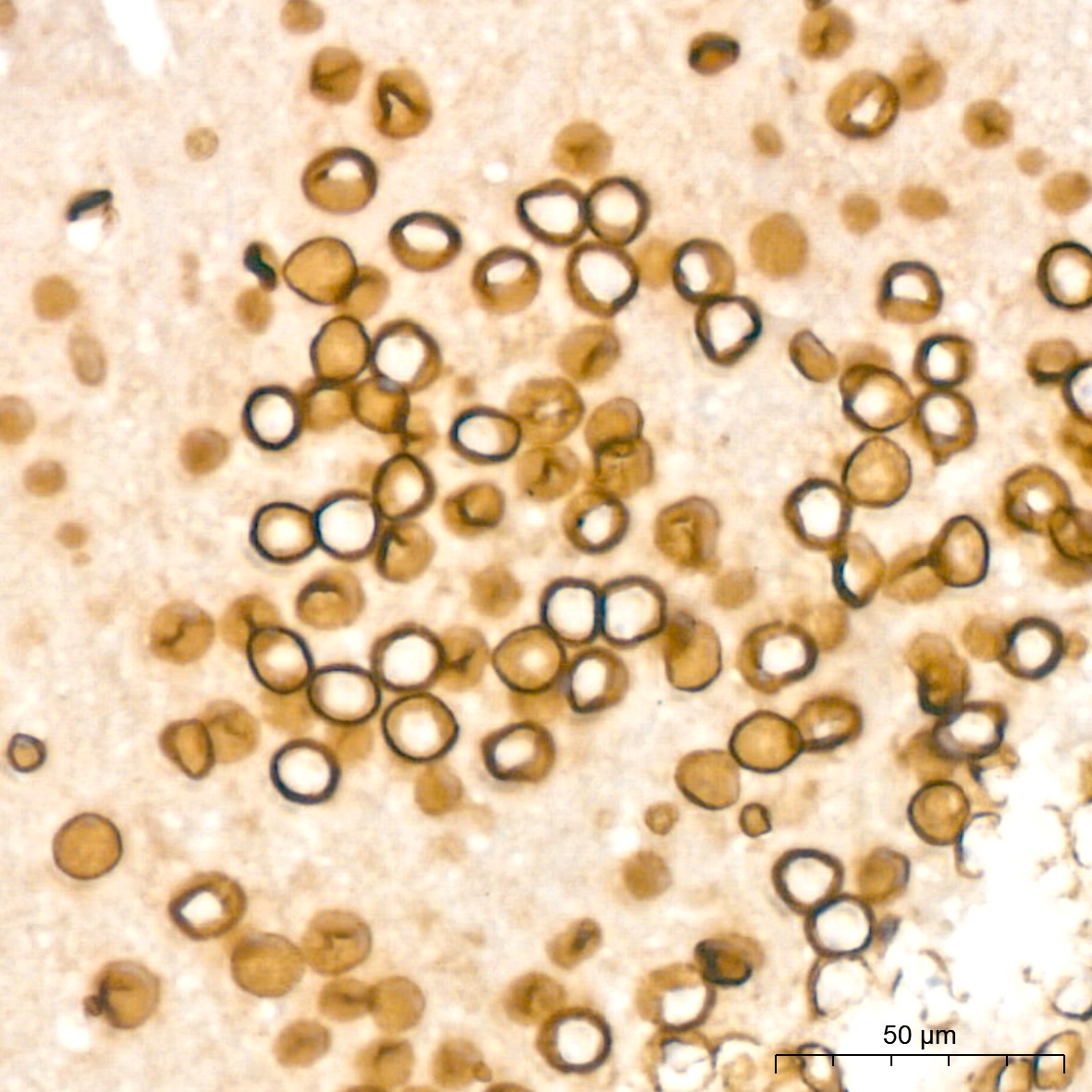
Immunohistochemistry analysis of paraffin-embedded Rat brain using Lamin B2 Rabbit mAb (CAB5001) at dilution of 1:100 (40x lens). Microwave antigen retrieval performed with 0.01M PBS Buffer (pH 7.2) prior to IHC staining.
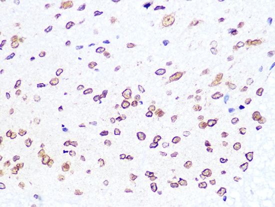
Immunohistochemistry analysis of paraffin-embedded Mouse spinal cord using Lamin B2 Rabbit mAb (CAB5001) at dilution of 1:100 (40x lens). Microwave antigen retrieval performed with 0.01M PBS Buffer (pH 7.2) prior to IHC staining.
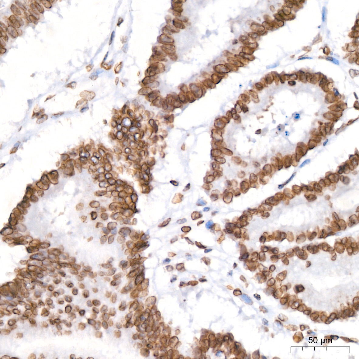
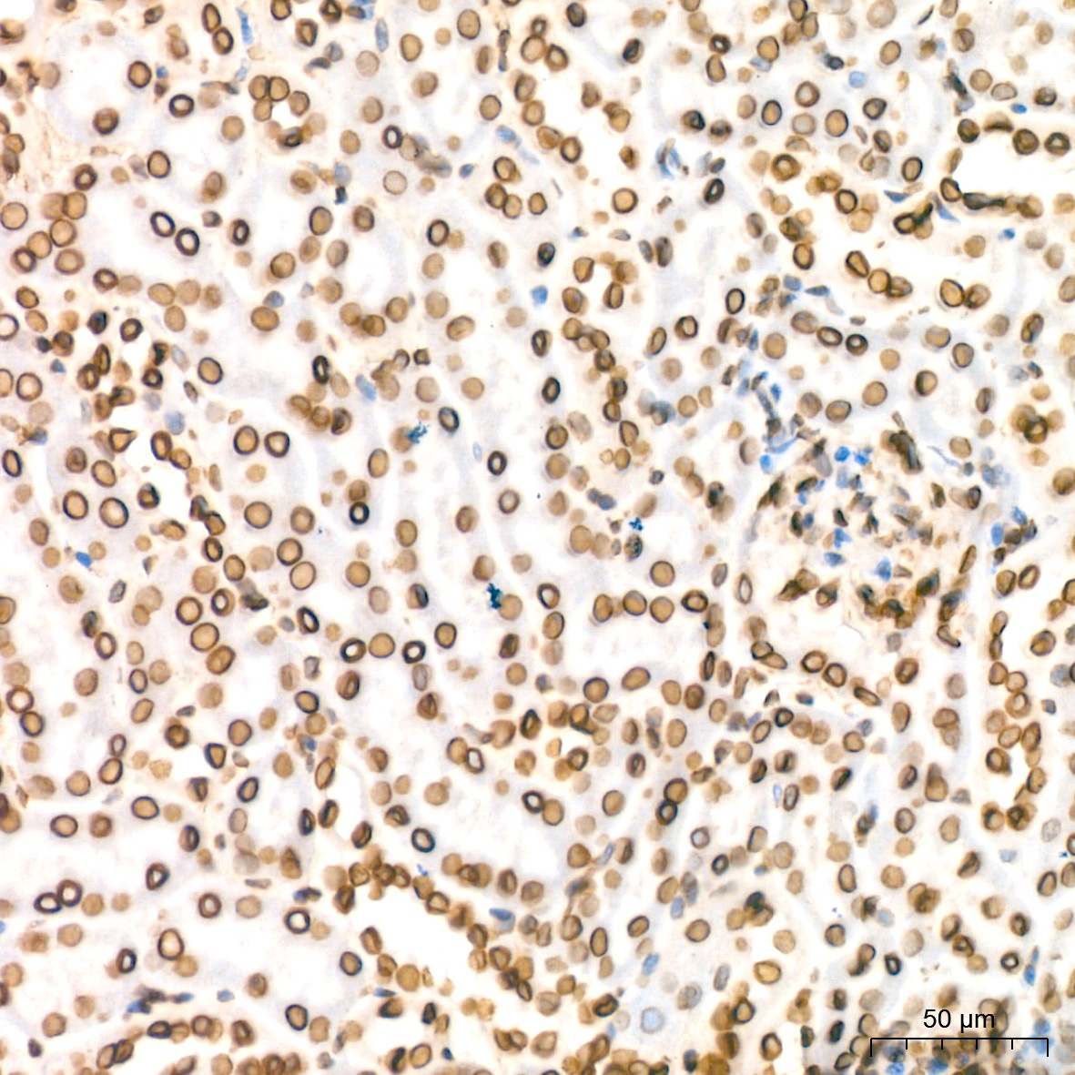
Immunohistochemistry analysis of paraffin-embedded Human colon carcinoma tissue using Lamin B2 Rabbit mAb (CAB22347) at a dilution of 1:200 (40x lens). High pressure antigen retrieval performed with 0.01M Citrate buffer (pH 6.0) prior to IHC staining.
Immunohistochemistry analysis of paraffin-embedded Mouse brain tissue using Lamin B2 Rabbit mAb (CAB22347) at a dilution of 1:200 (40x lens). High pressure antigen retrieval performed with 0.01M Citrate buffer (pH 6.0) prior to IHC staining.
Immunohistochemistry analysis of paraffin-embedded Mouse kidney tissue using Lamin B2 Rabbit mAb (CAB22347) at a dilution of 1:200 (40x lens). High pressure antigen retrieval performed with 0.01M Citrate buffer (pH 6.0) prior to IHC staining.
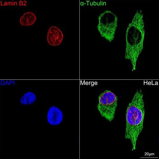
Confocal imaging of HeLa cells using Lamin B2 Rabbit mAb (CAB5001, dilution 1:100) (Red). The cells were counterstained with α-Tubulin Rabbit mAb (AC049, dilution 1:100) (Green). DAPI was used for nuclear staining (blue). Objective: 60x.