The LAMP2 Antibody (CAB1961) is a high-quality antibody developed for reliable detection and analysis of target proteins. LAMP2 is a glycoprotein found in lysosomal membranes and plays a crucial role in lysosomal biogenesis and autophagy. This polyclonal antibody, produced in rabbits, displays high reactivity with human samples and is verified for use in Western blot applications. By binding to the LAMP2 protein, this antibody allows for accurate detection and analysis in a wide range of cell types. With its specificity and sensitivity, it is particularly suited for studies in cell biology, molecular biology, and disease research.
This antibody is validated for use in WB, IHC-P, IF/ICC, ELISA applications and has demonstrated reactivity against Human, Mouse samples.
Product Name:
LAMP2 Antibody
SKU:
CAB1961
Size:
20μL, 100μL
Reactivity:
Human, Mouse
Conjugate:
Unconjugated
Immunogen:
Recombinant protein (or fragment).This information is considered to be commercially sensitive.
Recommended starting concentration is 1 μg/mL. Please optimize the concentration based on your specific assay requirements.
Synonyms:
DND, LAMPB, CD107b, LAMP-2, LGP-96, LGP110, LAMP2
Positive Sample:
SK-MEL-28, HeLa, Mouse kidney
Cellular Localization:
Cell Membrane, Endosome Membrane, Lysosome Membrane, Single-Pass Type I Membrane Protein.
Calculated MW:
45kDa
Observed MW:
100-130kDa
The protein encoded by this gene is a member of a family of membrane glycoproteins. This glycoprotein provides selectins with carbohydrate ligands. It may play a role in tumor cell metastasis. It may also function in the protection, maintenance, and adhesion of the lysosome. Alternative splicing of this gene results in multiple transcript variants encoding distinct proteins.
Purification Method
Affinity purification
Gene ID
3920
RRID
AB_2763987
Buffer Information
Store at -20℃. Avoid freeze / thaw cycles. Buffer: PBS containing 50% glycerol, preserved with proclin300 or sodium azide,pH7.3.
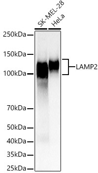
Western blot analysis of various lysates, using LAMP2 Rabbit pAb (CAB1961) at 1:2000 dilution. Secondary antibody: HRP-conjugated Goat anti-Rabbit IgG (H+L) (CABS014) at 1:10000 dilution. Lysates/proteins: 25μg per lane. Blocking buffer: 3% nonfat dry milk in TBST. Detection: ECL Basic Kit (AbGn00020). Exposure time: 3s.
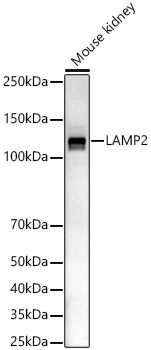
Western blot analysis of lysates from Mouse kidney, using LAMP2 Rabbit pAb (CAB1961) at 1:2000 dilution. Secondary antibody: HRP-conjugated Goat anti-Rabbit IgG (H+L) (CABS014) at 1:10000 dilution. Lysates/proteins: 25μg per lane. Blocking buffer: 3% nonfat dry milk in TBST. Detection: ECL Basic Kit (AbGn00020). Exposure time: 180s.
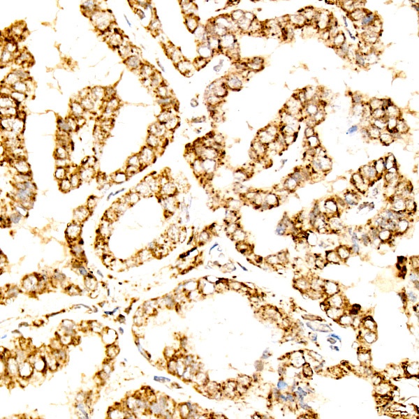
Immunohistochemistry analysis of paraffin-embedded Human thyroid cancer using LAMP2 Rabbit pAb (CAB1961) at dilution of 1:300 (40x lens). High pressure antigen retrieval performed with 0.01M Citrate buffer (pH 6.0) prior to IHC staining.
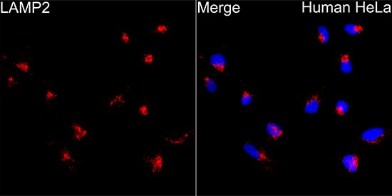
Immunofluorescence analysis of HeLa cells using LAMP2 Rabbit pAb (CAB1961) at dilution of 1:300 (40x lens). Secondary antibody: Cy3-conjugated Goat anti-Rabbit IgG (H+L) (CABS007) at 1:500 dilution. Blue: DAPI for nuclear staining.