The LHCGR Polyclonal Antibody (CAB24884) is a high-quality antibody developed for reliable detection and analysis of target proteins. This antibody, generated in rabbits, exhibits high specificity and sensitivity for detecting LHCGR in a variety of sample types, including cell lysates and tissue sections. Validated for use in Western blot and immunohistochemistry applications, this antibody allows for precise and accurate analysis of LHCGR expression in different cell types and tissues.The LHCGR, a G protein-coupled receptor, plays a critical role in the regulation of reproductive hormones and processes, such as ovulation and steroidogenesis. Dysregulation of LHCGR function has been implicated in various reproductive disorders, including infertility and ovarian hyperstimulation syndrome.
This antibody is validated for use in WB, ELISA, IF-P applications and has demonstrated reactivity against Mouse samples.
Product Name:
LHCGR Polyclonal Antibody
SKU:
CAB24884
Size:
20μL, 100μL
Reactivity:
Mouse
Conjugate:
Unconjugated
Immunogen:
Synthetic peptide. This information is considered to be commercially sensitive.
This gene encodes the receptor for both luteinizing hormone and choriogonadotropin. This receptor belongs to the G-protein coupled receptor 1 family, and its activity is mediated by G proteins which activate adenylate cyclase. Mutations in this gene result in disorders of male secondary sexual character development, including familial male precocious puberty, also known as testotoxicosis, hypogonadotropic hypogonadism, Leydig cell adenoma with precocious puberty, and male pseudohermaphtoditism with Leydig cell hypoplasia.
Purification Method
Affinity purification
Gene ID
3973
Buffer Information
Store at -20℃. Avoid freeze / thaw cycles. Buffer: PBS containing 50% glycerol, preserved with proclin300 or sodium azide, pH 7.3.
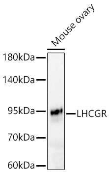
Western blot analysis of lysates from Mouse ovary using LHCGR Rabbit pAb (CAB24884) at 1:2000 dilution. Secondary antibody: HRP-conjugated Goat anti-Rabbit IgG (H+L) (CABS014) at 1:10000 dilution. Lysates/proteins: 25 μg per lane. Blocking buffer: 3% nonfat dry milk in TBST. Detection: ECL Basic Kit (AbGn00020). Exposure time: 60s.
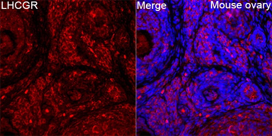
Immunofluorescence analysis of Mouse ovary tissue using LHCGR Rabbit pAb (CAB24884) at a dilution of 1:200 (40x lens). Secondary antibody: Cy3-conjugated Goat anti-Rabbit IgG (H+L)(CABS007) at 1:500 dilution. Blue: DAPI for nuclear staining. High pressure antigen retrieval performed with 0.01M Citrate Buffer (pH 6.0) prior to IF staining.
")
")


 at 1:10000 dilution. Lysates/proteins: 25ug per lane. Blocking buffer: 3% nonfat dry milk in TBST. Detection: ECL Enhanced Kit. Exposure time: 300s.")

")
. Blue: DAPI for nuclear staining.")
 at 1:10000 dilution. Lysates/proteins: 25ug per lane. Blocking buffer: 3% nonfat dry milk in TBST. Detection: ECL Basic Kit. Exposure time: 180s.")