The LSM14A Antibody (CAB16682) is a high-quality antibody developed for reliable detection and analysis of target proteins. This antibody, produced in rabbits, exhibits high specificity and sensitivity when detecting LSM14A in human samples, making it ideal for applications such as Western blotting.LSM14A plays a crucial role in post-transcriptional gene regulation, specifically in mRNA decay pathways. By targeting LSM14A, researchers can gain insights into the mechanisms that govern gene expression and RNA metabolism.
This antibody is validated for use in WB, IF/ICC, ELISA applications and has demonstrated reactivity against Human, Mouse, Rat samples.
Product Name:
LSM14A Antibody
SKU:
CAB16682
Size:
20μL, 100μL
Reactivity:
Human, Mouse, Rat
Conjugate:
Unconjugated
Immunogen:
Synthetic peptide. This information is considered to be commercially sensitive.
Recommended starting concentration is 1 μg/mL. Please optimize the concentration based on your specific assay requirements.
Synonyms:
RAP55, FAM61A, RAP55A, C19orf13, LSM14A
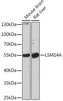
Positive Sample:
Mouse brain, Rat liver
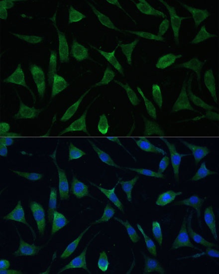
Cellular Localization:
Cytoplasm, P-Body.
Calculated MW:
51kDa
Observed MW:
50kDa
Sm-like proteins were identified in a variety of organisms based on sequence homology with the Sm protein family (see SNRPD2; 601061). Sm-like proteins contain the Sm sequence motif, which consists of 2 regions separated by a linker of variable length that folds as a loop. The Sm-like proteins are thought to form a stable heteromer present in tri-snRNP particles, which are important for pre-mRNA splicing.
Purification Method
Affinity purification
Gene ID
26065
RRID
AB_2770224
Buffer Information
Store at -20℃. Avoid freeze / thaw cycles. Buffer: PBS with 0.01% thimerosal,50% glycerol,pH7.3.
Western blot analysis of various lysates using LSM14A Rabbit pAb (CAB16682) at 1:1000 dilution. Secondary antibody: HRP-conjugated Goat anti-Rabbit IgG (H+L) (CABS014) at 1:10000 dilution. Lysates/proteins: 25μg per lane. Blocking buffer: 3% nonfat dry milk in TBST. Detection: ECL Enhanced Kit (AbGn00021). Exposure time: 90s.
Immunofluorescence analysis of L929 cells using LSM14A Rabbit pAb (CAB16682) at dilution of 1:100. Secondary antibody: Cy3-conjugated Goat anti-Rabbit IgG (H+L) (CABS007) at 1:500 dilution. Blue: DAPI for nuclear staining.