The [KO Validated] M6PR Antibody (CAB19907) is a high-quality antibody developed for reliable detection and analysis of target proteins. This rabbit polyclonal antibody is highly specific and sensitive, making it ideal for Western blot applications in various cell types.M6PR, or Mannose-6-Phosphate Receptor, plays a critical role in the sorting and delivery of lysosomal enzymes to their correct destinations within the cell. Dysregulation of M6PR expression and function has been implicated in various diseases, including lysosomal storage disorders and cancer.
This antibody is validated for use in WB, IHC-P, ELISA applications and has demonstrated reactivity against Human, Mouse, Rat samples.
Product Name:
[KO Validated] M6PR Antibody
SKU:
CAB19907
Size:
20μL, 100μL
Reactivity:
Human, Mouse, Rat
Conjugate:
Unconjugated
Immunogen:
Recombinant protein (or fragment).This information is considered to be commercially sensitive.
Recommended starting concentration is 1 μg/mL. Please optimize the concentration based on your specific assay requirements.
Synonyms:
SMPR, MPR46, CD-MPR, MPR 46, MPR-46, CD-M6PR, PR
Positive Sample:
293T, Mouse kidney
Cellular Localization:
Lysosome Membrane, Single-Pass Type I Membrane Protein.
Calculated MW:
31kDa
Observed MW:
31kDa
This gene encodes a member of the P-type lectin family. P-type lectins play a critical role in lysosome function through the specific transport of mannose-6-phosphate-containing acid hydrolases from the Golgi complex to lysosomes. The encoded protein functions as a homodimer and requires divalent cations for ligand binding. Alternatively spliced transcript variants encoding multiple isoforms have been observed for this gene. A pseudogene of this gene is located on the long arm of chromosome X.
Purification Method
Affinity purification
Gene ID
4074
RRID
AB_2862817
Buffer Information
Store at -20℃. Avoid freeze / thaw cycles. Buffer: PBS containing 50% glycerol, preserved with proclin300 or sodium azide, pH 7.3.
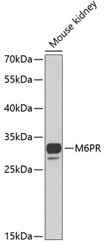
Western blot analysis of lysates from mouse kidney, using M6PR Rabbit pAb (CAB19907) at 1:1000 dilution. Secondary antibody: HRP-conjugated Goat anti-Rabbit IgG (H+L) (CABS014) at 1:10000 dilution. Lysates/proteins: 25μg per lane. Blocking buffer: 3% nonfat dry milk in TBST. Detection: ECL Basic Kit (AbGn00020). Exposure time: 40s.
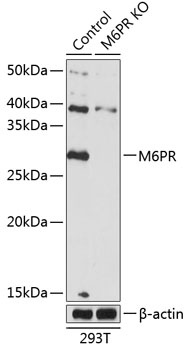
Western blot analysis of lysates from wild type (WT) and M6PR knockout (KO) 293T cells, using [KO Validated] M6PR Rabbit pAb (CAB19907) at 1:500 dilution. Secondary antibody: HRP-conjugated Goat anti-Rabbit IgG (H+L) (CABS014) at 1:10000 dilution. Lysates/proteins: 25μg per lane. Blocking buffer: 3% nonfat dry milk in TBST. Detection: ECL Basic Kit (AbGn00020). Exposure time: 1s.
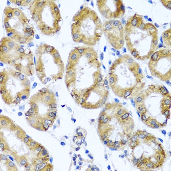
Immunohistochemistry analysis of paraffin-embedded Human stomach using M6PR Rabbit pAb (CAB19907) at dilution of 1:100 (40x lens). Microwave antigen retrieval performed with 0.01M PBS Buffer (pH 7.2) prior to IHC staining.
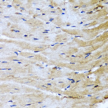
Immunohistochemistry analysis of paraffin-embedded Mouse heart using M6PR Rabbit pAb (CAB19907) at dilution of 1:100 (40x lens). Microwave antigen retrieval performed with 0.01M PBS Buffer (pH 7.2) prior to IHC staining.