The MAD1/MAD1L1 Monoclonal Antibody (CAB5098) is a high-quality antibody developed for reliable detection and analysis of target proteins. This antibody, produced using rabbit monoclonal technology, is highly specific for detecting MAD1 in human samples and has been validated for use in Western blot and immunofluorescence applications.MAD1 plays a crucial role in ensuring accurate chromosome segregation during cell division, making it a vital target for research in cancer biology and genetic disorders. By binding to MAD1, this antibody enables researchers to visualize and analyze the expression and localization of MAD1 in various cell types, providing valuable insights into its function and potential therapeutic implications.
This antibody is validated for use in WB, ELISA applications and has demonstrated reactivity against Human, Mouse samples.
Product Name:
MAD1/MAD1L1 Monoclonal Antibody
SKU:
CAB5098
Size:
20μL, 100μL
Reactivity:
Human, Mouse
Clone Number:
ARC1188
Conjugate:
Unconjugated
Immunogen:
Synthetic peptide. This information is considered to be commercially sensitive.
MAD1L1 is a component of the mitotic spindle-assembly checkpoint that prevents the onset of anaphase until all chromosome are properly aligned at the metaphase plate. MAD1L1 functions as a homodimer and interacts with MAD2L1. MAD1L1 may play a role in cell cycle control and tumor suppression. Alternative splicing results in multiple transcript variants.
Purification Method
Affinity purification
Gene ID
8379
RRID
AB_2863443
Buffer Information
Store at -20℃. Avoid freeze / thaw cycles. Buffer: PBS containing 50% glycerol and 0.05% BSA, preserved with proclin300 or sodium azide, pH 7.3.
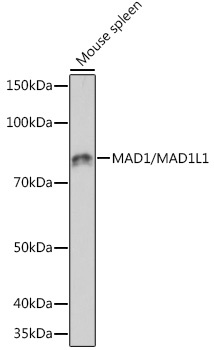
Western blot analysis of lysates from mouse spleen, using MAD1/MAD1L1 Rabbit mAb (CAB5098) at 1:1000 dilution. Secondary antibody: HRP-conjugated Goat anti-Rabbit IgG (H+L) (CABS014) at 1:10000 dilution. Lysates/proteins: 25 μg per lane. Blocking buffer: 3% nonfat dry milk in TBST. Detection: ECL Basic Kit (AbGn00020). Exposure time: 60 s.
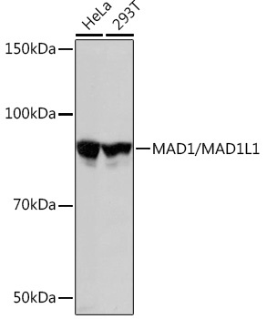
Western blot analysis of various lysates using MAD1/MAD1L1 Rabbit mAb (CAB5098) at 1:3000 dilution. Secondary antibody: HRP-conjugated Goat anti-Rabbit IgG (H+L) (CABS014) at 1:10000 dilution. Lysates/proteins: 25μg per lane. Blocking buffer: 3% nonfat dry milk in TBST. Detection: ECL Basic Kit (AbGn00020). Exposure time: 3s.