The MAD1/MAD1L1 Antibody (CAB1153) is a high-quality antibody developed for reliable detection and analysis of target proteins. This antibody, generated in rabbits, exhibits high reactivity with human samples and has been validated for use in Western blot applications. By binding specifically to the MAD1L1 protein, the antibody enables precise detection and analysis in various cell types, making it an essential component for studies in molecular biology and cancer research.MAD1L1, also known as mitotic spindle assembly checkpoint protein MAD1, plays a crucial role in ensuring proper chromosome segregation during cell division.
This antibody is validated for use in WB, IF/ICC, ELISA applications and has demonstrated reactivity against Human samples.
Product Name:
MAD1/MAD1L1 Antibody
SKU:
CAB1153
Size:
20μL, 100μL
Reactivity:
Human
Conjugate:
Unconjugated
Immunogen:
Recombinant protein (or fragment).This information is considered to be commercially sensitive.
MAD1L1 is a component of the mitotic spindle-assembly checkpoint that prevents the onset of anaphase until all chromosome are properly aligned at the metaphase plate. MAD1L1 functions as a homodimer and interacts with MAD2L1. MAD1L1 may play a role in cell cycle control and tumor suppression. Alternative splicing results in multiple transcript variants.
Purification Method
Affinity purification
Gene ID
8379
RRID
AB_2758595
Buffer Information
Store at -20℃. Avoid freeze / thaw cycles. Buffer: PBS containing 50% glycerol, preserved with proclin300 or sodium azide, pH 7.3.
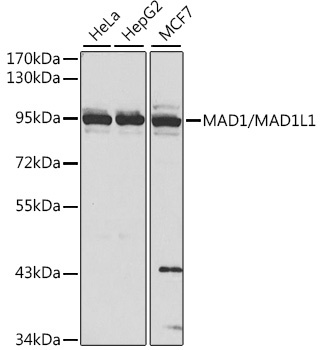
Western blot analysis of various lysates using MAD1/MAD1L1 Rabbit pAb (CAB1153) at 1:1000 dilution. Secondary antibody: HRP-conjugated Goat anti-Rabbit IgG (H+L) (CABS014) at 1:10000 dilution. Lysates/proteins: 25μg per lane. Blocking buffer: 3% nonfat dry milk in TBST. Detection: ECL Basic Kit (AbGn00020).
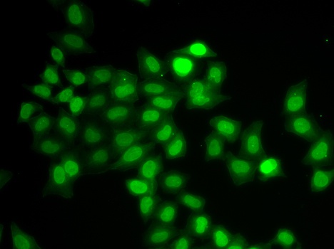
Immunofluorescence analysis of U2OS cells using MAD1/MAD1L1 Rabbit pAb (CAB1153).Secondary antibody: Cy3-conjugated Goat anti-Rabbit IgG (H+L) (CABS007) at 1:500 dilution.