The [KO Validated] LC3B Antibody (CAB11282) is a high-quality antibody developed for reliable detection and analysis of target proteins. Raised in rabbits, this antibody is highly specific to human samples and has been validated for use in Western blot applications. By binding to the MAP1LC3B protein, this antibody enables the accurate detection and analysis of autophagy processes in various cell types, making it an essential tool for studies in cell biology, cancer research, and neurodegenerative diseases.
This antibody is validated for use in WB, IHC-P, IF/ICC, ELISA applications and has demonstrated reactivity against Human, Mouse, Rat samples.
Product Name:
[KO Validated] LC3B Antibody
SKU:
CAB11282
Size:
20μL, 100μL
Reactivity:
Human, Mouse, Rat
Conjugate:
Unconjugated
Immunogen:
Recombinant protein (or fragment).This information is considered to be commercially sensitive.
The product of this gene is a subunit of neuronal microtubule-associated MAP1A and MAP1B proteins, which are involved in microtubule assembly and important for neurogenesis. Studies on the rat homolog implicate a role for this gene in autophagy, a process that involves the bulk degradation of cytoplasmic component.
Purification Method
Affinity purification
Gene ID
81631
RRID
AB_2861539
Buffer Information
Store at -20℃. Avoid freeze / thaw cycles. Buffer: PBS containing 50% glycerol, preserved with proclin300 or sodium azide, pH 7.3.
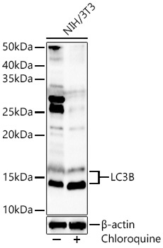
Western blot analysis of lysates from NIH/3T3 cells using LC3B Rabbit pAb (CAB11282) at 1:1000 dilution. NIH/3T3 cells were treated with Chloroquine (50 μM) at 37℃ for 20 hours. Secondary antibody: HRP-conjugated Goat anti-Rabbit IgG (H+L) (CABS014) at 1:10000 dilution. Lysates/proteins: 25 μg per lane. Blocking buffer: 3% nonfat dry milk in TBST. Detection: ECL Basic Kit (AbGn00020). Exposure time: 60s.
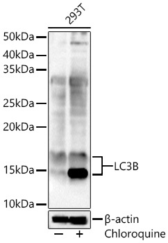
Western blot analysis of lysates from 293T cells using LC3B Rabbit pAb (CAB11282) at 1:1000 dilution. 293T cells were treated with Chloroquine (50 μM) at 37℃ for 20 hours. Secondary antibody: HRP-conjugated Goat anti-Rabbit IgG (H+L) (CABS014) at 1:10000 dilution. Lysates/proteins: 25 μg per lane. Blocking buffer: 3% nonfat dry milk in TBST. Detection: ECL Basic Kit (AbGn00020). Exposure time: 60s.
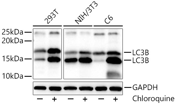
Western blot analysis of various lysates using LC3B Rabbit pAb (CAB11282)at 1:1000 dilution incubated overnight at 4℃. 293T,NIH/3T3,C6 cells were treated with Chloroquine at 37℃ for 48 hours. Secondary antibody: HRP-conjugated Goat anti-Rabbit IgG (H+L) (CABS014) at 1:10000 dilution. Lysates/proteins: 30 μg per lane. Blocking buffer: 3% nonfat dry milk in TBST. Detection: ECL Basic Kit (AbGn00020). Exposure time: 30s.
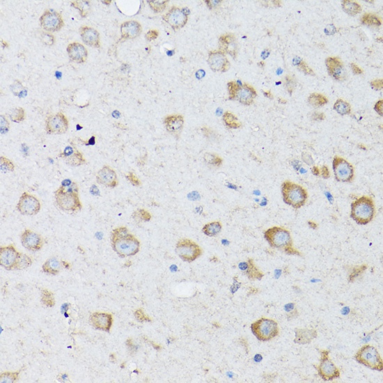
Immunohistochemistry analysis of paraffin-embedded Mouse brain using LC3B Rabbit pAb (CAB11282) at dilution of 1:100 (40x lens). High pressure antigen retrieval performed with 0.01M Citrate buffer (pH 6.0) prior to IHC staining.
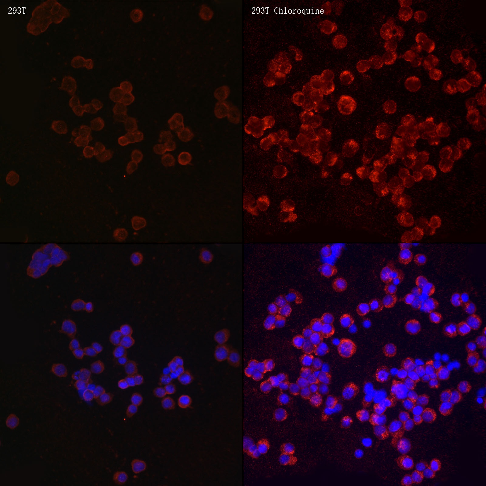
Immunofluorescence analysis of 293T and 293T Chloroquine cells using LC3B Rabbit pAb (CAB11282) at dilution of 1:100 (40x lens). Secondary antibody: Cy3-conjugated Goat anti-Rabbit IgG (H+L) (CABS007) at 1:500 dilution. Blue: DAPI for nuclear staining.