The [KO Validated] LC3B Antibody (CAB7198) is a high-quality antibody developed for reliable detection and analysis of target proteins. This antibody, produced in rabbits, has high specificity and sensitivity for detecting the MAP1LC3B protein in human samples, making it ideal for use in Western blot and immunofluorescence applications.MAP1LC3B, a key component of the autophagy pathway, is involved in the formation of autophagosomes which engulf and degrade cellular components, including damaged organelles and proteins. Dysregulation of autophagy has been linked to various diseases, including neurodegenerative disorders, cancer, and metabolic conditions.
This antibody is validated for use in WB, IHC-P, ELISA, IF-P applications and has demonstrated reactivity against Human, Mouse, Rat samples.
Product Name:
[KO Validated] LC3B Antibody
SKU:
CAB7198
Size:
20μL, 100μL
Reactivity:
Human, Mouse, Rat
Conjugate:
Unconjugated
Immunogen:
Synthetic peptide. This information is considered to be commercially sensitive.
The product of this gene is a subunit of neuronal microtubule-associated MAP1A and MAP1B proteins, which are involved in microtubule assembly and important for neurogenesis. Studies on the rat homolog implicate a role for this gene in autophagy, a process that involves the bulk degradation of cytoplasmic component.
Purification Method
Affinity purification
Gene ID
81631
RRID
AB_2863546
Buffer Information
Store at -20℃. Avoid freeze / thaw cycles. Buffer: PBS containing 50% glycerol, preserved with proclin300 or sodium azide, pH 7.3.
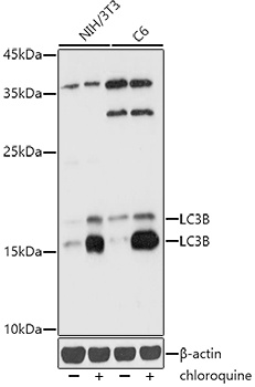
Western blot analysis of various lysates using [KD Validated] LC3B Rabbit pAb (CAB7198) at 1:1000 dilution. NIH/3T3 and C6 cells were treated with Chloroquine (50 μM) at 37℃ for 20 hours. Secondary antibody: HRP-conjugated Goat anti-Rabbit IgG (H+L) (CABS014) at 1:10000 dilution. Lysates/proteins: 25μg per lane. Blocking buffer: 3% nonfat dry milk in TBST. Detection: ECL Basic Kit (AbGn00020). Exposure time: 1s.
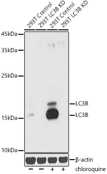
Western blot analysis of lysates from wild type (WT) and LC3B knockdown (KD) 293T,293T+chloroquine cells, using [KD Validated] LC3B Rabbit pAb (CAB7198) at 1:1000 dilution. 293T cells were treated with Chloroquine (50 μM) at 37℃ for 20 hours. Secondary antibody: HRP-conjugated Goat anti-Rabbit IgG (H+L) (CABS014) at 1:10000 dilution. Lysates/proteins: 25μg per lane. Blocking buffer: 3% nonfat dry milk in TBST. Detection: ECL Basic Kit (AbGn00020). Exposure time: 10s.
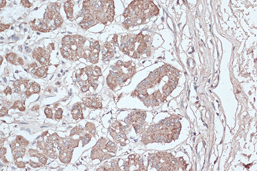
Immunohistochemistry analysis of paraffin-embedded Human breast cancer using [KD Validated] LC3B Rabbit pAb (CAB7198) at dilution of 1:100 (40x lens). Microwave antigen retrieval performed with 0.01M PBS Buffer (pH 7.2) prior to IHC staining.
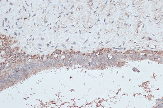
Immunohistochemistry analysis of paraffin-embedded Human stomach using [KD Validated] LC3B Rabbit pAb (CAB7198) at dilution of 1:100 (40x lens). Microwave antigen retrieval performed with 0.01M PBS Buffer (pH 7.2) prior to IHC staining.