The MCAT Antibody (CAB15822) is a high-quality antibody developed for reliable detection and analysis of target proteins. This antibody, developed in rabbits, exhibits high reactivity with human samples and has been validated for use in Western blot applications. By binding specifically to the MCAT protein, this antibody allows for precise detection and analysis in various cell types, making it an essential component in studies related to metabolism, lipid synthesis, and mitochondrial function.MCAT is a critical enzyme that catalyzes the conversion of malonyl-CoA to malonyl-ACP during fatty acid biosynthesis in mitochondria.
This antibody is validated for use in WB, ELISA applications and has demonstrated reactivity against Human samples.
Product Name:
MCAT Antibody
SKU:
CAB15822
Size:
20μL, 100μL
Reactivity:
Human
Conjugate:
Unconjugated
Immunogen:
Recombinant protein (or fragment).This information is considered to be commercially sensitive.
Recommended starting concentration is 1 μg/mL. Please optimize the concentration based on your specific assay requirements.
Synonyms:
MT, MCT, MCT1, fabD, NET62, FASN2C, MCAT
Positive Sample:
LO2
Cellular Localization:
Mitochondrion.
Calculated MW:
43kDa
Observed MW:
43kDa
The protein encoded by this gene is found exclusively in the mitochondrion, where it catalyzes the transfer of a malonyl group from malonyl-CoA to the mitochondrial acyl carrier protein. The encoded protein may be part of a fatty acid synthase complex that is more like the type II prokaryotic and plastid complexes rather than the type I human cytosolic complex. Alternative splicing results in multiple transcript variants encoding different isoforms.
Purification Method
Affinity purification
Gene ID
27349
RRID
AB_2763246
Buffer Information
Store at -20℃. Avoid freeze / thaw cycles. Buffer: PBS with 0.01% thimerosal,50% glycerol,pH7.3.
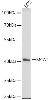
Western blot analysis of lysates from LO2 cells, using MCAT Rabbit pAb (CAB15822) at 1:1000 dilution. Secondary antibody: HRP-conjugated Goat anti-Rabbit IgG (H+L) (CABS014) at 1:10000 dilution. Lysates/proteins: 25μg per lane. Blocking buffer: 3% nonfat dry milk in TBST. Detection: ECL Basic Kit (AbGn00020). Exposure time: 90s.