The MEK1/MEK2 Antibody (CAB18117) is a high-quality antibody developed for reliable detection and analysis of target proteins. This antibody, generated in rabbits, is highly specific for human samples and has been validated for use in Western blot applications. By binding to MEK1 and MEK2, this antibody allows for the detection and analysis of these important signaling molecules in a variety of cell types.MEK1 and MEK2 are critical components of the MAPK/ERK pathway, which regulates cell growth, differentiation, and survival.
This antibody is validated for use in WB, ELISA applications and has demonstrated reactivity against Mouse, Rat samples.
Product Name:
MEK1/MEK2 Antibody
SKU:
CAB18117
Size:
20μL, 100μL
Reactivity:
Mouse, Rat
Conjugate:
Unconjugated
Immunogen:
Synthetic peptide. This information is considered to be commercially sensitive.
Recommended starting concentration is 1 μg/mL. Please optimize the concentration based on your specific assay requirements.
Synonyms:
MEK1/MEK2
Positive Sample:
mouse brain, mouse spleen, PC-12
Cellular Localization:
Cytosol, Early Endosome, Endoplasmic Reticulum, Focal Adhesion, Golgi Apparatus, Late Endosome, Microtubule Organizing Center, Mitochondrion, Nucleus, Plasma Membrane.
Calculated MW:
40kDa/43kDa/44kDa
Observed MW:
44kDa
The protein MEK1 and MEK2 are two members of the dual specificity protein kinase family, which act as mitogen-activated protein (MAP) kinase kinase. MAP kinases, also known as extracellular signal-regulated kinases (ERKs), act as an integration point for multiple biochemical signals. This protein kinase lies upstream of MAP kinases and stimulates the enzymatic activity of MAP kinases upon wide variety of extra- and intracellular signals. As an essential component of MAP kinase signal transduction pathway, this kinase is involved in many cellular processes such as proliferation, differentiation, transcription regulation and development.
Purification Method
Affinity purification
Gene ID
5604 5605
RRID
AB_2861909
Buffer Information
Store at -20℃. Avoid freeze / thaw cycles. Buffer: PBS containing 50% glycerol, preserved with proclin300 or sodium azide, pH 7.3.
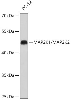
Western blot analysis of lysates from PC-12 cells, using MEK1/MEK2 Rabbit pAb (CAB18117) at 1:500 dilution. Secondary antibody: HRP-conjugated Goat anti-Rabbit IgG (H+L) (CABS014) at 1:10000 dilution. Lysates/proteins: 25μg per lane. Blocking buffer: 3% nonfat dry milk in TBST. Detection: ECL Basic Kit (AbGn00020). Exposure time: 30s.
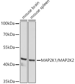
Western blot analysis of various lysates using MEK1/MEK2 Rabbit pAb (CAB18117) at 1:500 dilution. Secondary antibody: HRP-conjugated Goat anti-Rabbit IgG (H+L) (CABS014) at 1:10000 dilution. Lysates/proteins: 25μg per lane. Blocking buffer: 3% nonfat dry milk in TBST. Detection: ECL Basic Kit (AbGn00020). Exposure time: 60s.