The METTL16 Antibody (CAB15894) is a high-quality antibody developed for reliable detection and analysis of target proteins. This antibody, produced in rabbits, is highly specific to Mettl16 in human samples and has been validated for use in Western blot applications.Mettl16 is a methyltransferase enzyme that plays a crucial role in the methylation of RNA, particularly in the modification of transfer RNA (tRNA). Dysregulation of Mettl16 has been linked to various diseases, including cancer and neurological disorders.
This antibody is validated for use in WB, IHC-P, IF/ICC, IP, ELISA applications and has demonstrated reactivity against Human, Mouse, Rat samples.
Product Name:
METTL16 Antibody
SKU:
CAB15894
Size:
20μL, 100μL
Reactivity:
Human, Mouse, Rat
Conjugate:
Unconjugated
Immunogen:
Recombinant protein (or fragment).This information is considered to be commercially sensitive.
0.5μg-4μg antibody for 200μg-400μg extracts of whole cells
ELISA
Recommended starting concentration is 1 μg/mL. Please optimize the concentration based on your specific assay requirements.
Synonyms:
METT10D, METTL16
Positive Sample:
HCT 116, LNCaP
Cellular Localization:
Cytoplasm, Nucleus.
Calculated MW:
64kDa
Observed MW:
78kDa
Enables RNA binding activity and RNA methyltransferase activity. Involved in RNA modification and regulation of mRNA metabolic process. Located in cytoplasm and nucleus.
Purification Method
Affinity purification
Gene ID
79066
RRID
AB_2763325
Buffer Information
Store at -20℃. Avoid freeze / thaw cycles. Buffer: PBS with 0.09% Sodium azide,50% glycerol,pH7.3.
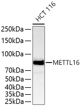
Western blot analysis of lysates from HCT 116 cells using METTL16 Rabbit pAb (CAB15894) at 1:5000 dilution incubated overnight at 4℃. Secondary antibody: HRP-conjugated Goat anti-Rabbit IgG (H+L) (CABS014) at 1:10000 dilution. Lysates/proteins: 25 μg per lane. Blocking buffer: 3% nonfat dry milk in TBST. Detection: ECL Basic Kit (AbGn00020). Exposure time: 45 s.
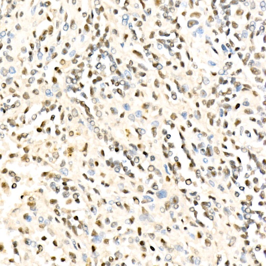
Immunohistochemistry analysis of paraffin-embedded Human spleen using METTL16 Rabbit pAb (CAB15894) at dilution of 1:100 (40x lens). High pressure antigen retrieval performed with 0.01M Citrate buffer (pH 6.0) prior to IHC staining.
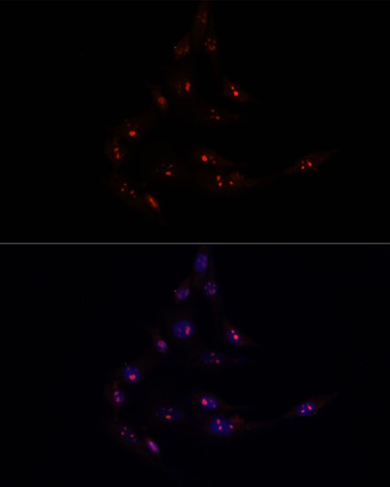
Immunofluorescence analysis of NIH/3T3 cells using METTL16 Rabbit pAb (CAB15894) at dilution of 1:100. Secondary antibody: Cy3-conjugated Goat anti-Rabbit IgG (H+L) (CABS007) at 1:500 dilution. Blue: DAPI for nuclear staining.
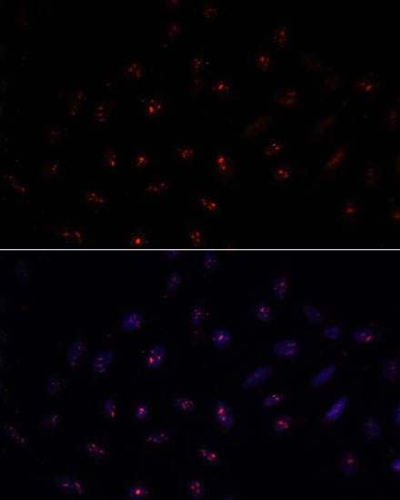
Immunofluorescence analysis of U-2 OS cells using METTL16 Rabbit pAb (CAB15894) at dilution of 1:100. Secondary antibody: Cy3-conjugated Goat anti-Rabbit IgG (H+L) (CABS007) at 1:500 dilution. Blue: DAPI for nuclear staining.