The METTL3 Antibody (CAB8370) is a high-quality antibody developed for reliable detection and analysis of target proteins. This antibody, produced in rabbits, exhibits high reactivity with human samples and has been validated for use in Western blot applications. By binding specifically to the Mettl3 protein, this antibody enables the detection and analysis of Mettl3 in a variety of cell types, making it well-suited for investigations in molecular biology and cancer research.Mettl3 is a key player in the process of mRNA methylation, which has been linked to various cellular functions including RNA stability, translation efficiency, and protein synthesis.
This antibody is validated for use in WB, IHC-P, IF/ICC, ELISA applications and has demonstrated reactivity against Human, Mouse, Rat samples.
Product Name:
METTL3 Antibody
SKU:
CAB8370
Size:
20μL, 100μL
Reactivity:
Human, Mouse, Rat
Conjugate:
Unconjugated
Immunogen:
Recombinant protein (or fragment).This information is considered to be commercially sensitive.
Recommended starting concentration is 1 μg/mL. Please optimize the concentration based on your specific assay requirements.
Synonyms:
M6A, IME4, Spo8, MT-A70, hMETTL3, METTL3
Positive Sample:
NIH/3T3, 293T, HeLa, Hep G2, Mouse testis
Cellular Localization:
Nucleus Speckle.
Calculated MW:
64kDa
Observed MW:
70kDa/75KDa/75kDa
This gene encodes the 70 kDa subunit of MT-A which is part of N6-adenosine-methyltransferase. This enzyme is involved in the posttranscriptional methylation of internal adenosine residues in eukaryotic mRNAs, forming N6-methyladenosine.
Purification Method
Affinity purification
Gene ID
56339
RRID
AB_2770344
Buffer Information
Store at -20℃. Avoid freeze / thaw cycles. Buffer: PBS containing 50% glycerol, preserved with proclin300 or sodium azide, pH 7.3.
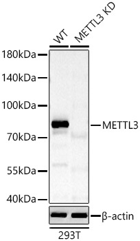
Western blot analysis of lysates from wild type (WT) and METTL3 knockdown (KD) 293T cells using METTL3 Rabbit pAb (CAB8370) at 1:1000 dilution incubated overnight at 4℃. Secondary antibody: HRP-conjugated Goat anti-Rabbit IgG (H+L) (CABS014) at 1:10000 dilution. Lysates/proteins: 25 μg per lane. Blocking buffer: 3% nonfat dry milk in TBST. Detection: ECL Basic Kit (AbGn00020). Exposure time: 30s.
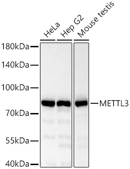
Western blot analysis of various lysates using METTL3 Rabbit pAb (CAB8370) at 1:1000 dilution incubated overnight at 4℃. Secondary antibody: HRP-conjugated Goat anti-Rabbit IgG (H+L) (CABS014) at 1:10000 dilution. Lysates/proteins: 25 μg per lane. Blocking buffer: 3% nonfat dry milk in TBST. Detection: ECL Basic Kit (AbGn00020). Exposure time: 30s.
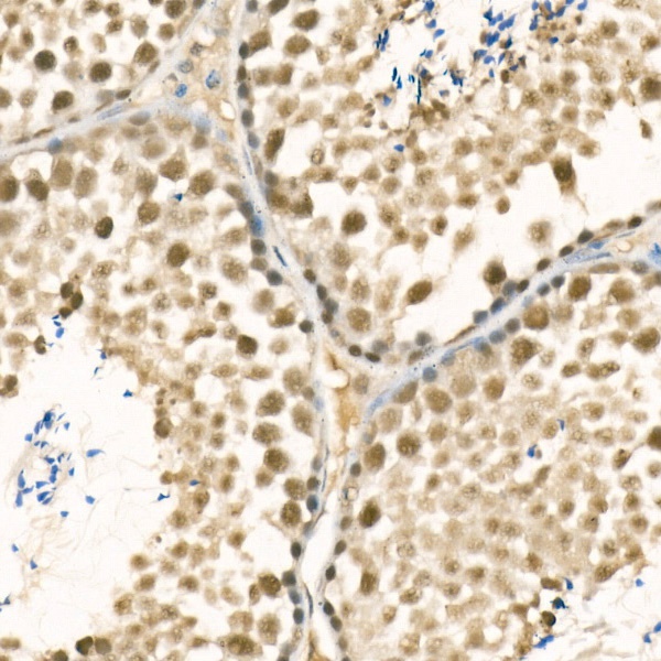
Immunohistochemistry analysis of paraffin-embedded Mouse testis using METTL3 Rabbit pAb (CAB8370) at dilution of 1:100 (40x lens). High pressure antigen retrieval performed with 0.01M Citrate buffer (pH 6.0) prior to IHC staining.
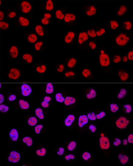
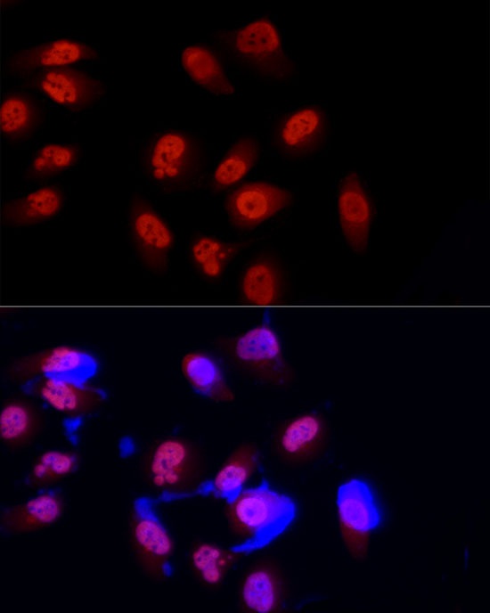
Confocal immunofluorescence analysis of Hela cells using METTL3 Rabbit pAb (CAB8370) at dilution of 1:200. Blue: DAPI for nuclear staining.
Immunofluorescence analysis of PC-3 cells using METTL3 Rabbit pAb (CAB8370) at dilution of 1:100 (40x lens). Secondary antibody: Cy3-conjugated Goat anti-Rabbit IgG (H+L) (CABS007) at 1:500 dilution. Blue: DAPI for nuclear staining.