The MT-ATP8 Antibody (CAB17890) is a high-quality antibody developed for reliable detection and analysis of target proteins. This antibody, raised in rabbits, exhibits high reactivity with human samples and has been validated for use in Western blot applications.MT-ATP8 is essential for the synthesis of ATP, the energy currency of the cell, and its dysfunction has been linked to various mitochondrial disorders and diseases. By binding specifically to the MT-ATP8 protein, this antibody enables accurate detection and analysis of this crucial mitochondrial component in a variety of cell types.
This antibody is validated for use in WB, IHC-P, IF/ICC, ELISA applications and has demonstrated reactivity against Human, Mouse, Rat samples.
Product Name:
MT-ATP8 Antibody
SKU:
CAB17890
Size:
20μL, 100μL
Reactivity:
Human, Mouse, Rat
Immunogen:
Synthetic peptide. This information is considered to be commercially sensitive.
Recommended starting concentration is 1 μg/mL. Please optimize the concentration based on your specific assay requirements.
Synonyms:
ATPase8, MTATP8, ATP8, MT-ATP8
Positive Sample:
HeLa treated with EB, Rat liver, Hep G2
Cellular Localization:
Mitochondrial Inner Membrane, Mitochondrial Proton-Transporting Atp Synthase Complex, Mitochondrial Proton-Transporting Atp Synthase Complex, Coupling Factor F(O).
Calculated MW:
8kDa
Observed MW:
8kDa
Contributes to proton-transporting ATP synthase activity, rotational mechanism. Involved in mitochondrial ATP synthesis coupled proton transport. Part of mitochondrial proton-transporting ATP synthase complex. Implicated in multiple sclerosis and urinary bladder cancer.
Purification Method
Affinity purification
Gene ID
4509
RRID
AB_2861745
Buffer Information
Store at -20℃. Avoid freeze / thaw cycles. Buffer: PBS containing 50% glycerol, preserved with proclin300 or sodium azide, pH 7.3.
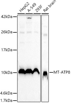
Western blot analysis of various lysates using MT-ATP8 Rabbit pAb (CAB17890) at 1:1000 dilution. Secondary antibody: HRP-conjugated Goat anti-Rabbit IgG (H+L) (CABS014) at 1:10000 dilution. Lysates/proteins: 25μg per lane. Blocking buffer: 3% nonfat dry milk in TBST. Detection: ECL Basic Kit (AbGn00020). Exposure time: 10s.
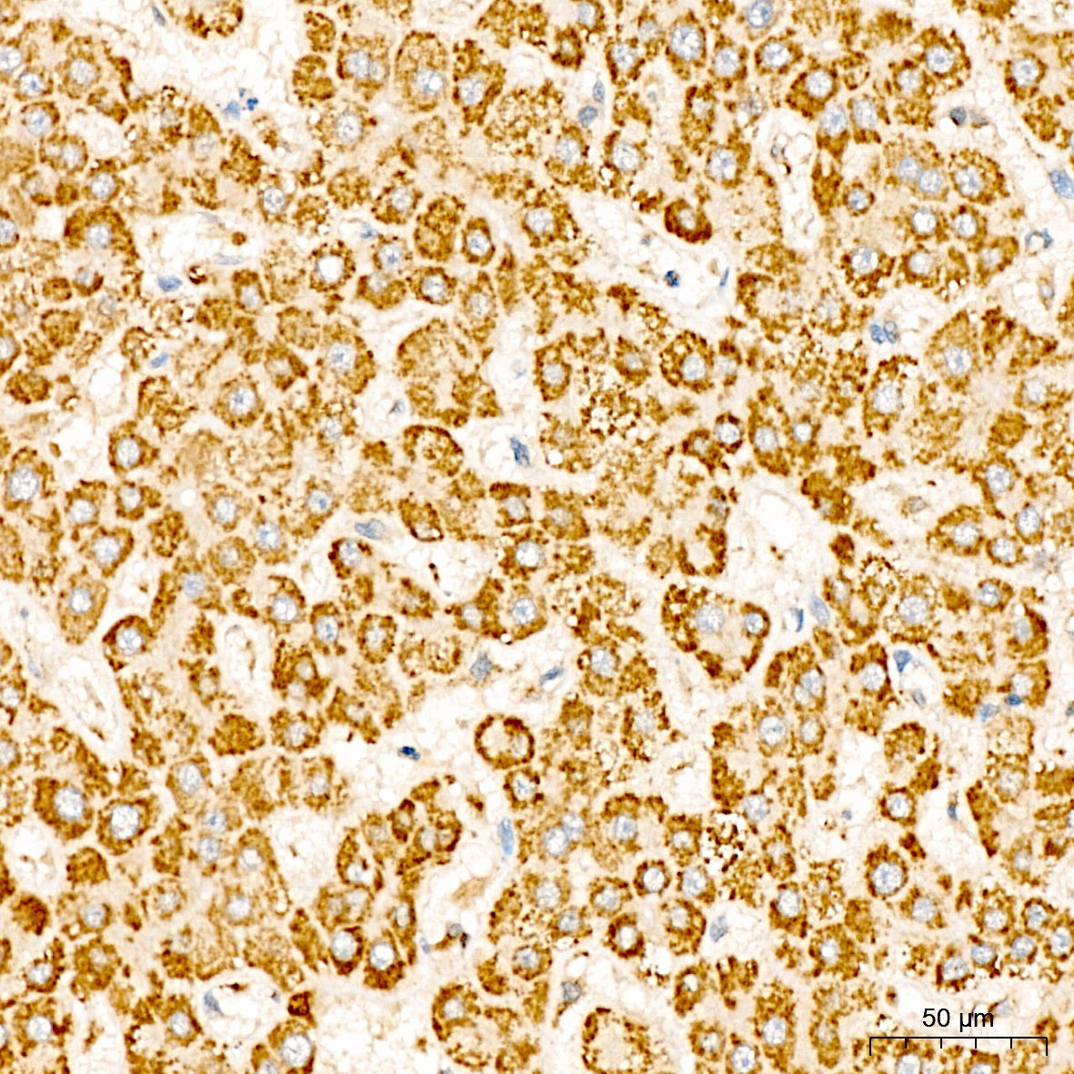
Immunohistochemistry analysis of paraffin-embedded Human liver tissue using MT-ATP8 Rabbit pAb (CAB17890) at a dilution of 1:100 (40x lens). High pressure antigen retrieval was performed with 0.01 M citrate buffer (pH 6.0) prior to IHC staining.
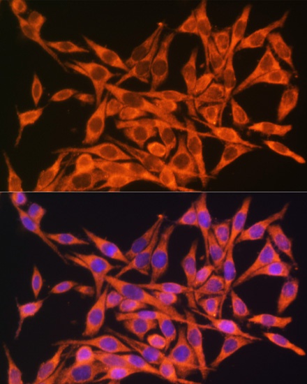
Immunofluorescence analysis of HeLa cells using MT-ATP8 Rabbit pAb (CAB17890) at dilution of 1:100. Secondary antibody: Cy3-conjugated Goat anti-Rabbit IgG (H+L) (CABS007) at 1:500 dilution. Blue: DAPI for nuclear staining.