The MT-ND2 Antibody (CAB17968) is a high-quality antibody developed for reliable detection and analysis of target proteins. This antibody, derived from rabbit serum, exhibits high specificity and sensitivity when detecting MT-ND2 in human samples, making it ideal for use in Western blot applications. By targeting the MT-ND2 protein, this antibody allows for the accurate identification and analysis of mitochondrial function in various cell types.The MT-ND2 protein is involved in the electron transport chain within mitochondria, playing a crucial role in energy production and cellular metabolism. Dysregulation of MT-ND2 has been linked to a variety of diseases, including neurodegenerative disorders and metabolic conditions.
This antibody is validated for use in WB, IHC-P, IF/ICC, ELISA applications and has demonstrated reactivity against Human, Mouse, Rat samples.
Product Name:
MT-ND2 Antibody
SKU:
CAB17968
Size:
20μL, 100μL
Reactivity:
Human, Mouse, Rat
Immunogen:
Synthetic peptide. This information is considered to be commercially sensitive.
Recommended starting concentration is 1 μg/mL. Please optimize the concentration based on your specific assay requirements.
Synonyms:
MTND2, ND2, MT-ND2
Positive Sample:
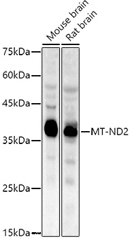
Mouse brain, Rat brain
Cellular Localization:
Mitochondrial Inner Membrane, Mitochondrial Respiratory Chain Complex I.
Calculated MW:
39kDa
Observed MW:
39kDa
Enables NADH dehydrogenase (ubiquinone) activity. Involved in mitochondrial electron transport, NADH to ubiquinone and mitochondrial respiratory chain complex I assembly. Part of mitochondrial respiratory chain complex I. Implicated in Leber hereditary optic neuropathy; multiple sclerosis; myocardial infarction; neurodegenerative disease (multiple); and urinary bladder cancer.
Purification Method
Affinity purification
Gene ID
4536
RRID
AB_2861770
Buffer Information
Store at -20℃. Avoid freeze / thaw cycles. Buffer: PBS with 0.09% Sodium azide,50% glycerol,pH7.3.
Western blot analysis of various lysates using MT-ND2 Rabbit pAb (CAB17968) at 1:1000 dilution. Secondary antibody: HRP-conjugated Goat anti-Rabbit IgG (H+L) (CABS014) at 1:10000 dilution. Lysates/proteins: 25μg per lane. Blocking buffer: 3% nonfat dry milk in TBST. Detection: ECL Enhanced Kit (AbGn00021). Exposure time: 90s.
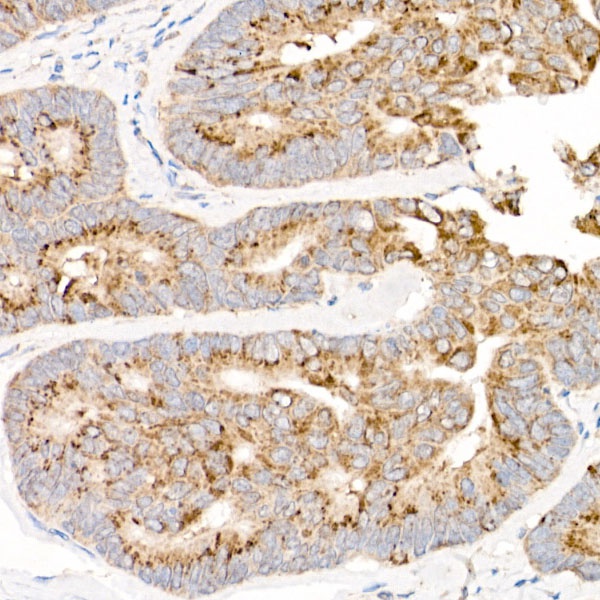
Immunohistochemistry analysis of paraffin-embedded Human colon carcinoma using MT-ND2 Rabbit pAb (CAB17968) at dilution of 1:50 (40x lens). High pressure antigen retrieval performed with 0.01M Citrate buffer (pH 6.0) prior to IHC staining.