The MTA1 Antibody (CAB16085) is a high-quality antibody developed for reliable detection and analysis of target proteins. This antibody, raised in rabbits, exhibits high reactivity with human samples and is validated for use in Western blot applications.By targeting the MTA1 protein, this antibody enables the detection and analysis of MTA1 expression in different cell types, making it an essential tool for investigating the role of MTA1 in cancer and other diseases.
This antibody is validated for use in WB, ELISA applications and has demonstrated reactivity against Human, Mouse samples.
Product Name:
MTA1 Antibody
SKU:
CAB16085
Size:
20μL, 100μL
Reactivity:
Human, Mouse
Conjugate:
Unconjugated
Immunogen:
Synthetic peptide. This information is considered to be commercially sensitive.
This gene encodes a protein that was identified in a screen for genes expressed in metastatic cells, specifically, mammary adenocarcinoma cell lines. Expression of this gene has been correlated with the metastatic potential of at least two types of carcinomas although it is also expressed in many normal tissues. The role it plays in metastasis is unclear. It was initially thought to be the 70kD component of a nucleosome remodeling deacetylase complex, NuRD, but it is more likely that this component is a different but very similar protein. These two proteins are so closely related, though, that they share the same types of domains. These domains include two DNA binding domains, a dimerization domain, and a domain commonly found in proteins that methylate DNA. The profile and activity of this gene product suggest that it is involved in regulating transcription and that this may be accomplished by chromatin remodeling. Two transcript variants encoding different isoforms have been found for this gene.
Purification Method
Affinity purification
Gene ID
9112
RRID
AB_2763526
Buffer Information
Store at -20℃. Avoid freeze / thaw cycles. Buffer: PBS containing 50% glycerol, preserved with proclin300 or sodium azide, pH 7.3.
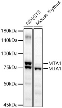
Western blot analysis of various lysates using MTA1 Rabbit pAb (CAB16085) at 1:2000 dilution. Secondary antibody: HRP-conjugated Goat anti-Rabbit IgG (H+L) (CABS014) at 1:10000 dilution. Lysates/proteins: 25μg per lane. Blocking buffer: 3% nonfat dry milk in TBST. Detection: ECL Basic Kit (AbGn00020). Exposure time: 10s.