The MTCO2 Monoclonal Antibody (CAB3843) is a high-quality antibody developed for reliable detection and analysis of target proteins. This antibody, raised in rabbits, exhibits high reactivity with human samples and is validated for use in various applications, including Western blot and immunohistochemistry.The MTCO2 protein plays a crucial role in cellular respiration by facilitating the transfer of electrons within the mitochondrial complex IV. Dysregulation of MTCO2 has been implicated in various metabolic disorders, neurodegenerative diseases, and cancer. Therefore, the MTCO2 Rabbit Monoclonal Antibody is an invaluable tool for studying mitochondrial function and exploring potential therapeutic targets in these pathologies.
This antibody is validated for use in WB, IHC-P, IF/ICC, ELISA, IF-P applications and has demonstrated reactivity against Human samples.
Product Name:
MTCO2 Monoclonal Antibody
SKU:
CAB3843
Size:
20μL, 100μL
Reactivity:
Human
Clone Number:
ARC0844
Conjugate:
Unconjugated
Immunogen:
Synthetic peptide. This information is considered to be commercially sensitive.
Contributes to cytochrome-c oxidase activity. Predicted to be involved in mitochondrial electron transport, cytochrome c to oxygen and positive regulation of vasoconstriction. Located in mitochondrial inner membrane. Part of respiratory chain complex IV. Biomarker of Huntington's disease and stomach cancer.
Purification Method
Affinity purification
Gene ID
4513
RRID
AB_2863148
Buffer Information
Store at -20℃. Avoid freeze / thaw cycles. Buffer: PBS containing 50% glycerol and 0.05% BSA, preserved with proclin300 or sodium azide, pH 7.3.
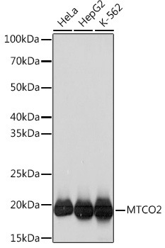
Western blot analysis of various lysates using MTCO2 Rabbit mAb (CAB3843) at 1:1000 dilution. Secondary antibody: HRP-conjugated Goat anti-Rabbit IgG (H+L) (CABS014) at 1:10000 dilution. Lysates/proteins: 25μg per lane. Blocking buffer: 3% nonfat dry milk in TBST. Detection: ECL Basic Kit (AbGn00020). Exposure time: 3s.
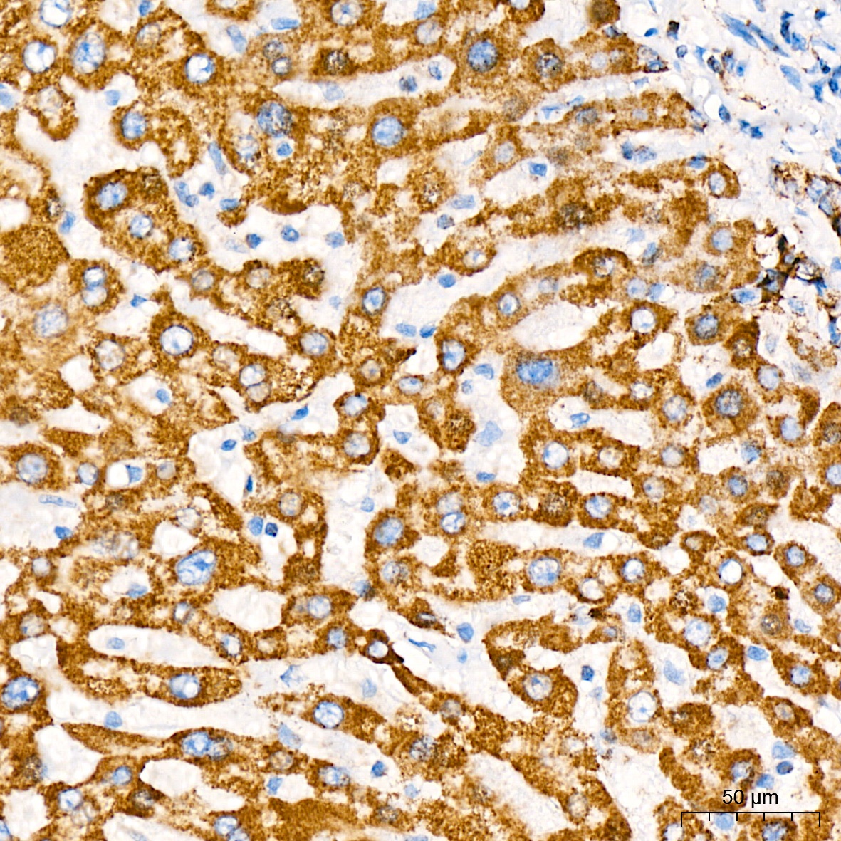
Immunohistochemistry analysis of paraffin-embedded Human liver tissue using MTCO2 Rabbit mAb (CAB3843) at a dilution of 1:200 (40x lens). High pressure antigen retrieval was performed with 0.01 M citrate buffer (pH 6.0) prior to IHC staining.
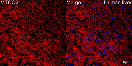
Confocal imaging of paraffin-embedded Human liver tissue using MTCO2 Rabbit mAb (CAB3843, dilution 1:200) followed by a further incubation with Cy3 Goat Anti-Rabbit IgG (H+L) (CABS007,dilution 1:500) (Red). DAPI was used for nuclear staining (Blue). Objective: 40x. Perform high pressure antigen retrieval with 0.01 M citrate buffer (pH 6.0) prior to IF staining.
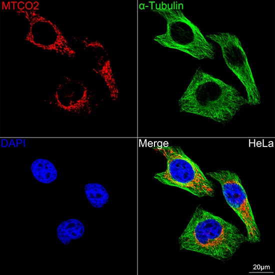
Confocal imaging of HeLa cells using MTCO2 Rabbit mAb (CAB3843, dilution 1:200) followed by a further incubation with Cy3 Goat Anti-Rabbit IgG (H+L) (CABS007, dilution 1:500) (Red). The cells were counterstained with α-Tubulin Mouse mAb (AC012, dilution 1:400) followed by incubation with ABflo® 488-conjugated Goat Anti-Mouse IgG (H+L) Ab (CABS076, dilution 1:500) (Green). DAPI was used for nuclear staining (Blue). Objective: 100x.