The NAPSA Polyclonal Antibody (CAB24485) is a high-quality antibody developed for reliable detection and analysis of target proteins. This antibody, derived from rabbit serum, demonstrates high reactivity with human samples and is validated for use in Western blot applications. By targeting the NAPSA protein, researchers can accurately detect and analyze its expression in various cell types, making it an essential tool for studies in immunology and cancer research.NAPSA, also known as aspartyl aminopeptidase, plays a key role in immune function by regulating inflammation and promoting immune balance.
This antibody is validated for use in WB, ELISA applications and has demonstrated reactivity against Rat samples.
Product Name:
NAPSA Polyclonal Antibody
SKU:
CAB24485
Size:
20μL, 100μL
Reactivity:
Rat
Conjugate:
Unconjugated
Immunogen:
Recombinant protein (or fragment).This information is considered to be commercially sensitive.
Recommended starting concentration is 1 μg/mL. Please optimize the concentration based on your specific assay requirements.
Synonyms:
NAPSA, KAP, Kdap, NAP1, NAPA, SNAPA, napsin-A
Positive Sample:
Rat lung
Cellular Localization:
Secreted.
Calculated MW:
45kDa
Observed MW:
38-55kDa
This gene encodes a member of the peptidase A1 family of aspartic proteases. The encoded preproprotein is proteolytically processed to generate an activation peptide and the mature protease. The activation peptides of aspartic proteinases function as inhibitors of the protease active site. These peptide segments, or pro-parts, are deemed important for correct folding, targeting, and control of the activation of aspartic proteinase zymogens. The encoded protease may play a role in the proteolytic processing of pulmonary surfactant protein B in the lung and may function in protein catabolism in the renal proximal tubules. This gene has been described as a marker for lung adenocarcinoma and renal cell carcinoma.
Purification Method
Affinity purification
Gene ID
9476
Buffer Information
Store at -20℃. Avoid freeze / thaw cycles. Buffer: PBS containing 50% glycerol, preserved with proclin300 or sodium azide, pH 7.3.
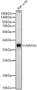
Western blot analysis of lysates from Rat lung, using NAPSA Rabbit pAb (CAB24485) at 1:2000 dilution. Secondary antibody: HRP-conjugated Goat anti-Rabbit IgG (H+L) (CABS014) at 1:10000 dilution. Lysates/proteins: 25μg per lane. Blocking buffer: 3% nonfat dry milk in TBST. Detection: ECL Basic Kit (AbGn00020). Exposure time: 30s.
 at 1:2000 dilution. Secondary antibody: HRP Goat Anti-Rabbit IgG (H+L) at 1:10000 dilution. Lysates/proteins: 25ug per lane. Blocking buffer: 3% nonfat dry milk in TBST.")
 at 1:2000 dilution. Secondary antibody: HRP Goat Anti-Rabbit IgG (H+L) at 1:10000 dilution. Lysates/proteins: 25ug per lane. Blocking buffer: 3% nonfat dry milk in TBST.")


 at 1:10000 dilution. Lysates/proteins: 25ug per lane. Blocking buffer: 3% nonfat dry milk in TBST. Detection: ECL Enhanced Kit. Exposure time: 300s.")

")
. Blue: DAPI for nuclear staining.")
 at 1:10000 dilution. Lysates/proteins: 25ug per lane. Blocking buffer: 3% nonfat dry milk in TBST. Detection: ECL Basic Kit. Exposure time: 180s.")