The NDUFS7 Polyclonal Antibody (CAB24466) is a high-quality antibody developed for reliable detection and analysis of target proteins. This antibody, produced in rabbits, exhibits high specificity and sensitivity towards human samples, making it suitable for Western blot applications. By binding to NDUFs7, this antibody enables the detection and analysis of this crucial protein in various cell types, facilitating research in bioenergetics and mitochondrial function.NDUFs7 is essential for the proper functioning of mitochondrial complex I, the largest enzyme complex in the respiratory chain responsible for generating ATP. Dysregulation of complex I activity has been linked to various mitochondrial disorders, neurodegenerative diseases, and cancer.
This antibody is validated for use in WB, ELISA applications and has demonstrated reactivity against Human samples.
Product Name:
NDUFS7 Polyclonal Antibody
SKU:
CAB24466
Size:
20μL, 100μL
Reactivity:
Human
Conjugate:
Unconjugated
Immunogen:
Synthetic peptide. This information is considered to be commercially sensitive.
This gene encodes a protein that is a subunit of one of the complexes that forms the mitochondrial respiratory chain. This protein is one of over 40 subunits found in complex I, the nicotinamide adenine dinucleotide (NADH):ubiquinone oxidoreductase. This complex functions in the transfer of electrons from NADH to the respiratory chain, and ubiquinone is believed to be the immediate electron acceptor for the enzyme. Mutations in this gene cause Leigh syndrome due to mitochondrial complex I deficiency, a severe neurological disorder that results in bilaterally symmetrical necrotic lesions in subcortical brain regions.
Purification Method
Affinity purification
Gene ID
374291
Buffer Information
Store at -20℃. Avoid freeze / thaw cycles. Buffer: PBS containing 50% glycerol, preserved with proclin300 or sodium azide, pH 7.3.
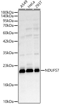
Western blot analysis of various lysates, using NDUFS7 Rabbit pAb (CAB24466) at 1:2000 dilution. Secondary antibody: HRP-conjugated Goat anti-Rabbit IgG (H+L) (CABS014) at 1:10000 dilution. Lysates/proteins: 25μg per lane. Blocking buffer: 3% nonfat dry milk in TBST. Detection: ECL Basic Kit (AbGn00020). Exposure time: 45s.
 at 1:2000 dilution. Secondary antibody: HRP Goat Anti-Rabbit IgG (H+L) at 1:10000 dilution. Lysates/proteins: 25ug per lane. Blocking buffer: 3% nonfat dry milk in TBST.")
 at 1:2000 dilution. Secondary antibody: HRP Goat Anti-Rabbit IgG (H+L) at 1:10000 dilution. Lysates/proteins: 25ug per lane. Blocking buffer: 3% nonfat dry milk in TBST.")

 at 1:10000 dilution. Lysates/proteins: 25ug per lane. Blocking buffer: 3% nonfat dry milk in TBST. Detection: ECL Enhanced Kit. Exposure time: 300s.")

")
. Blue: DAPI for nuclear staining.")
 at 1:10000 dilution. Lysates/proteins: 25ug per lane. Blocking buffer: 3% nonfat dry milk in TBST. Detection: ECL Basic Kit. Exposure time: 180s.")
 variety Zhonghua 11, using OsNAC4 antibody at 1:1000 dilution. Secondary antibody: HRP Goat Anti-Rabbit IgG (H+L) at 1:10000 dilution. Lysates/proteins: 25ug per lane. Blocking buffer: 3% nonfat dry milk in TBST. Detection: ECL Enhanced Kit. Exposure time: 30s.")