The NEK2 Monoclonal Antibody (CAB6811) is a high-quality antibody developed for reliable detection and analysis of target proteins. This antibody, developed using rabbit monoclonal technology, exhibits high specificity and sensitivity towards NEK2 in human samples, making it ideal for applications such as Western blotting.NEK2 plays a key role in ensuring accurate chromosome segregation during cell division, making it a crucial player in maintaining genomic stability. Dysregulation of NEK2 has been implicated in various cancers, making it a promising therapeutic target for cancer treatment.
This antibody is validated for use in WB, ELISA applications and has demonstrated reactivity against Human, Mouse, Rat samples.
Product Name:
NEK2 Monoclonal Antibody
SKU:
CAB6811
Size:
20μL, 100μL
Reactivity:
Human, Mouse, Rat
Clone Number:
ARC1418
Conjugate:
Unconjugated
Immunogen:
Synthetic peptide. This information is considered to be commercially sensitive.
Recommended starting concentration is 1 μg/mL. Please optimize the concentration based on your specific assay requirements.
Synonyms:
NLK1, RP67, NEK2A, HsPK21, PPP1R111, NEK2
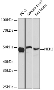
Positive Sample:
PC-3, Mouse testis, Rat testis
Cellular Localization:
Chromosome, Cytoplasm, Nucleus, Centromere, Centromere, Centrosome, Cytoskeleton, Kinetochore, Microtubule Organizing Center, Nucleolus, Spindle Pole.
Calculated MW:
52kDa
Observed MW:
50kDa
This gene encodes a serine/threonine-protein kinase that is involved in mitotic regulation. This protein is localized to the centrosome, and undetectable during G1 phase, but accumulates progressively throughout the S phase, reaching maximal levels in late G2 phase. Alternatively spliced transcript variants encoding different isoforms with distinct C-termini have been noted for this gene.
Purification Method
Affinity purification
Gene ID
4751
RRID
AB_2863536
Buffer Information
Store at -20℃. Avoid freeze / thaw cycles. Buffer: PBS containing 50% glycerol and 0.05% BSA, preserved with proclin300 or sodium azide, pH 7.3.
Western blot analysis of various lysates using NEK2 Rabbit mAb (CAB6811) at 1:1000 dilution. Secondary antibody: HRP-conjugated Goat anti-Rabbit IgG (H+L) (CABS014) at 1:10000 dilution. Lysates/proteins: 25μg per lane. Blocking buffer: 3% nonfat dry milk in TBST. Detection: ECL Basic Kit (AbGn00020). Exposure time: 30s.