The NONO/p54nrb Monoclonal Antibody (CAB3800) is a high-quality antibody developed for reliable detection and analysis of target proteins. This gene encodes an RNA-binding protein which plays various roles in the nucleus, including transcriptional regulation and RNA splicing. A rearrangement between this gene and the transcription factor E3 gene has been observed in papillary renal cell carcinoma. Alternatively spliced transcript variants have been described. Pseudogenes exist on Chromosomes 2 and 16. RRID AB_2863142 Gene ID 4841 Swiss Prot Synonym P54; NMT55; NRB54; MRXS34; P54NRB; PPP1R114; NONO/p54nrb
This antibody is validated for use in WB, IHC-P, IF/ICC, ELISA applications and has demonstrated reactivity against Human, Mouse, Rat samples.
Product Name:
NONO/p54nrb Monoclonal Antibody
SKU:
CAB3800
Size:
100μL, 20μL
Reactivity:
Human, Mouse, Rat
Clone Number:
ARC0836
Conjugate:
Unconjugated
Immunogen:
Synthetic peptide. This information is considered to be commercially sensitive.
Tested Applications:
WBIHC-PIF/ICCELISA
Recommended Dilution:
WB
1:1000 - 1:4000
IHC-P
1:500 - 1:2000
IF
/
ICC
1:100 - 1:1000
ELISA
Recommended starting concentration is 1 μg/mL. Please optimize the concentration based on your specific assay requirements.
This gene encodes an RNA-binding protein which plays various roles in the nucleus, including transcriptional regulation and RNA splicing. A rearrangement between this gene and the transcription factor E3 gene has been observed in papillary renal cell carcinoma. Alternatively spliced transcript variants have been described. Pseudogenes exist on Chromosomes 2 and 16. RRID AB_2863142 Gene ID 4841 Swiss Prot Synonym P54; NMT55; NRB54; MRXS34; P54NRB; PPP1R114; NONO/p54nrb
Purification Method:
Affinity purification
Gene ID:
4841
RRID:
AB_2863142
Buffer Information:
Store at -20℃. Avoid freeze / thaw cycles. Buffer: PBS containing 50% glycerol and 0.05% BSA, preserved with proclin300 or sodium azide, pH 7.3.
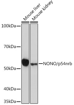
Western blot analysis of various lysates using NONO/p54nrb Rabbit mAb (CAB3800) at 1:1000 dilution. Secondary antibody: HRP-conjugated Goat anti-Rabbit IgG (H+L) (AS014) at 1:10000 dilution. Lysates/proteins: 25μg per lane. Blocking buffer: 3% nonfat dry milk in TBST. Detection: ECL Basic Kit (AbGn00020). Exposure time: 3min.
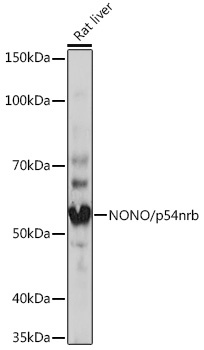
Western blot analysis of lysates from Rat liver, using NONO/p54nrb Rabbit mAb (CAB3800) at 1:1000 dilution. Secondary antibody: HRP-conjugated Goat anti-Rabbit IgG (H+L) (AS014) at 1:10000 dilution. Lysates/proteins: 25μg per lane. Blocking buffer: 3% nonfat dry milk in TBST. Detection: ECL Basic Kit (AbGn00020). Exposure time: 3min.
Immunohistochemistry analysis of paraffin-embedded Human tonsil tissue using NONO/p54nrb Rabbit mAb (CAB3800) at a dilution of 1:500 (40x lens). High pressure antigen retrieval performed with 0.01M Tris-EDTA Buffer (pH 9.0) prior to IHC staining.
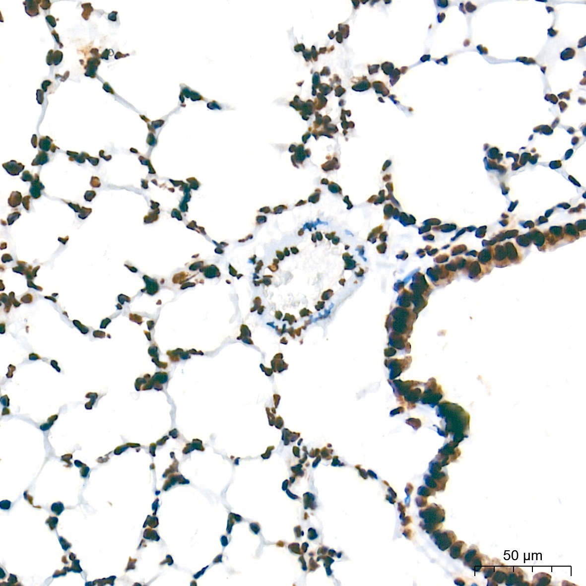
Immunohistochemistry analysis of paraffin-embedded Mouse lung tissue using NONO/p54nrb Rabbit mAb (CAB3800) at a dilution of 1:500 (40x lens). High pressure antigen retrieval performed with 0.01M Tris-EDTA Buffer (pH 9.0) prior to IHC staining.
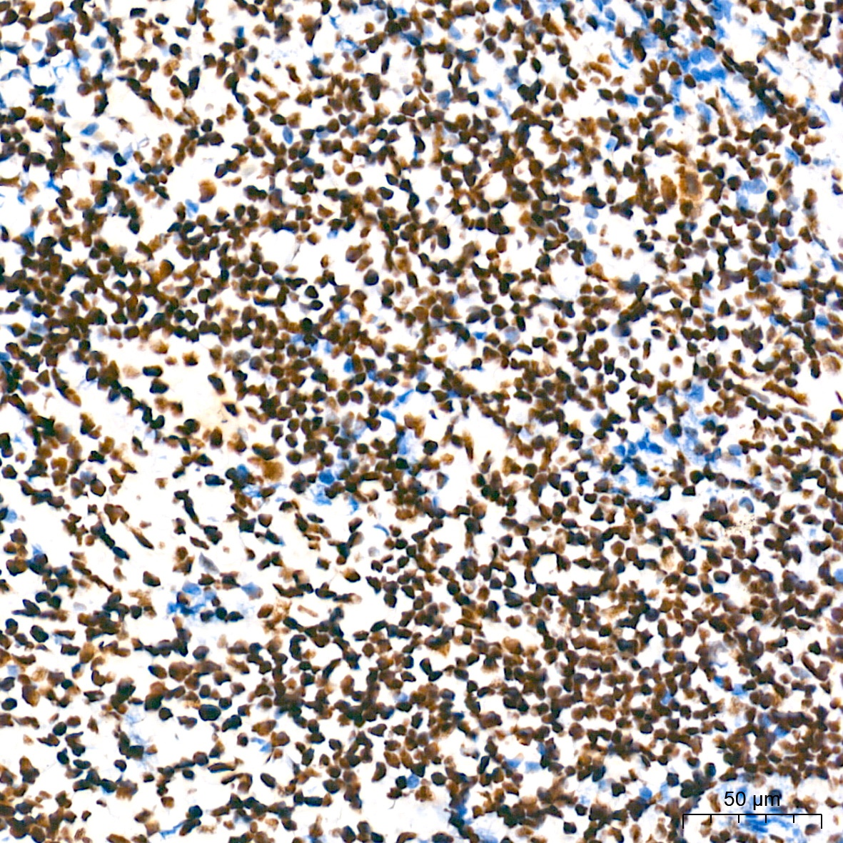
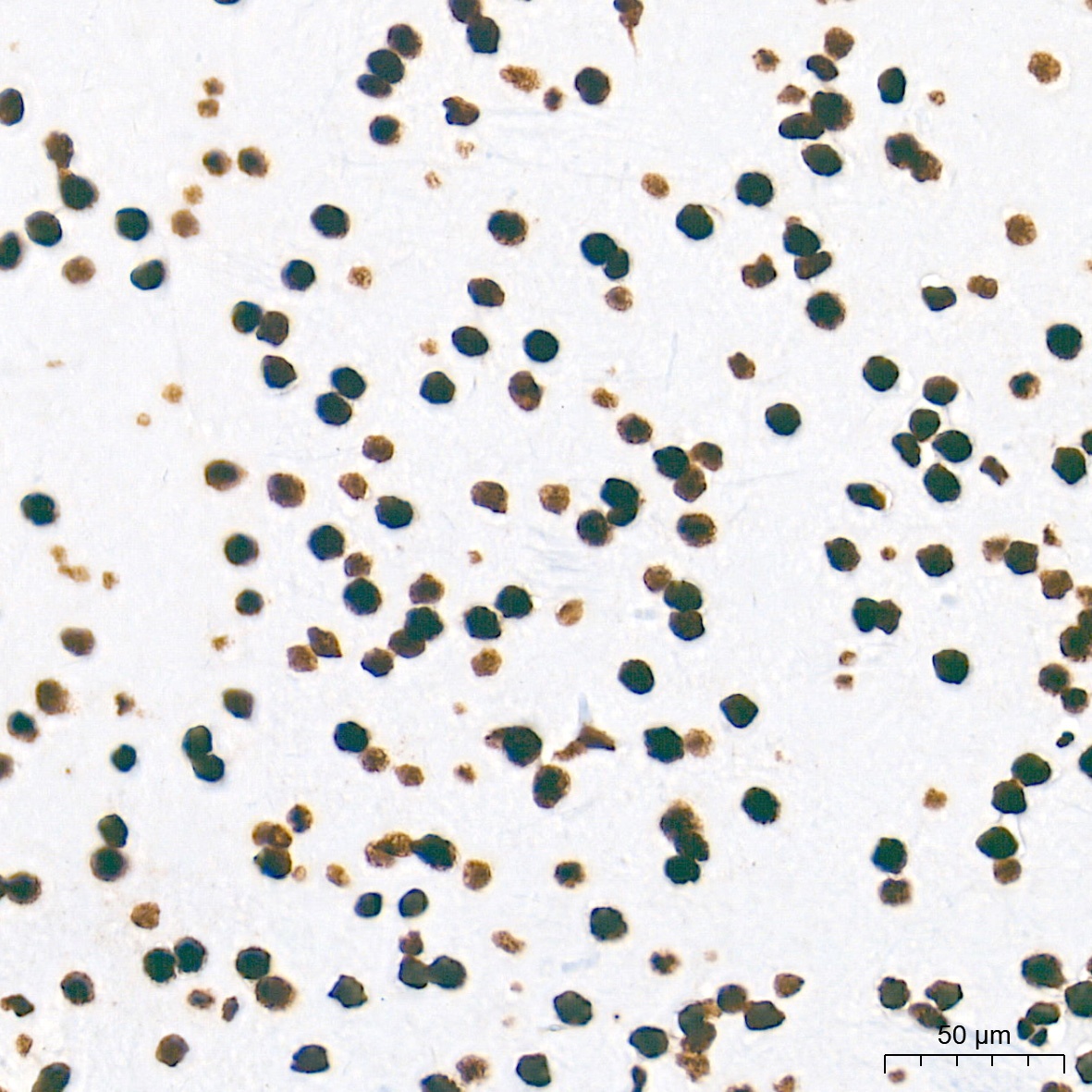
Immunohistochemistry analysis of paraffin-embedded Rat brain tissue using NONO/p54nrb Rabbit mAb (CAB3800) at a dilution of 1:500 (40x lens). High pressure antigen retrieval performed with 0.01M Tris-EDTA Buffer (pH 9.0) prior to IHC staining.
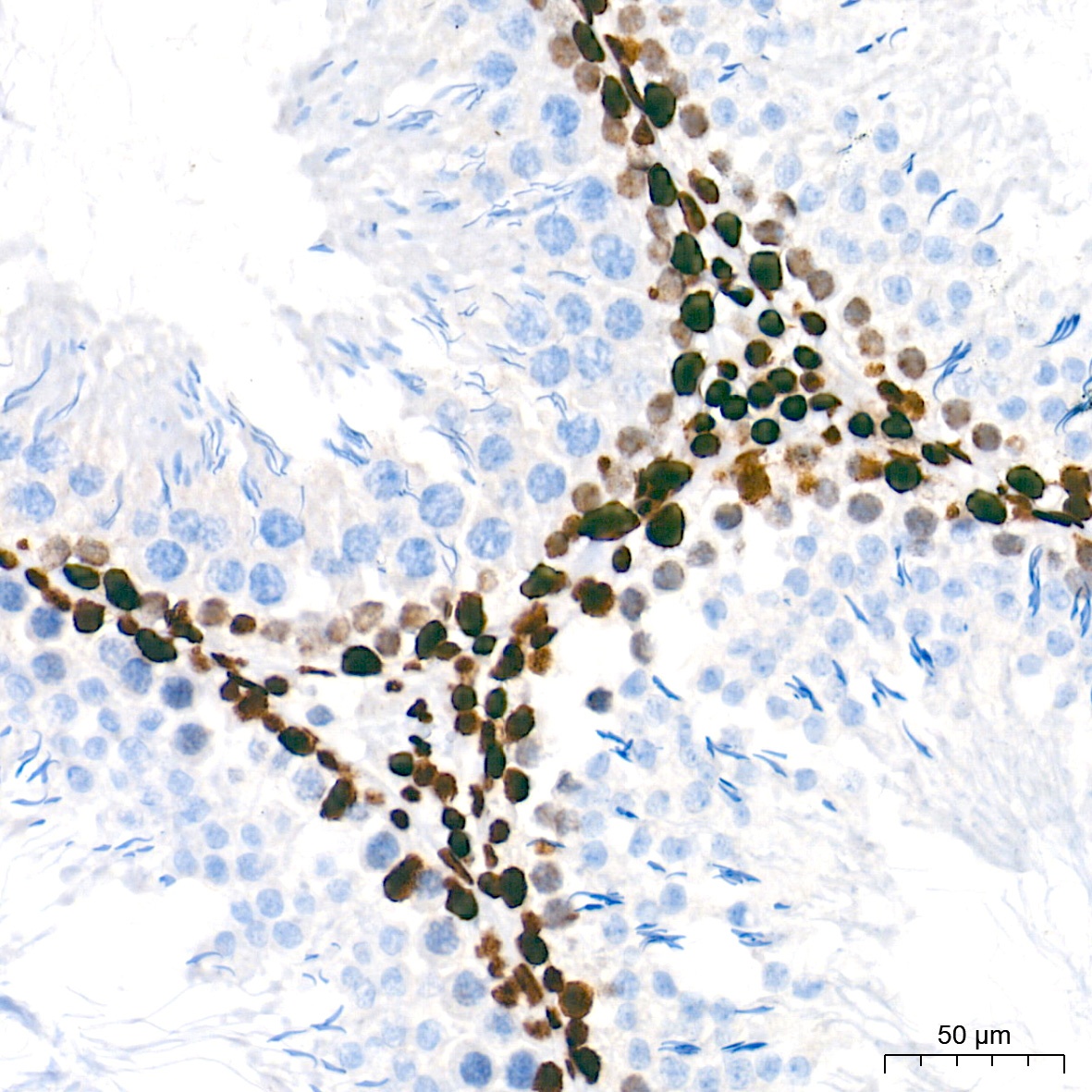
Immunohistochemistry analysis of paraffin-embedded Rat testis tissue using NONO/p54nrb Rabbit mAb (CAB3800) at a dilution of 1:500 (40x lens). High pressure antigen retrieval performed with 0.01M Tris-EDTA Buffer (pH 9.0) prior to IHC staining.
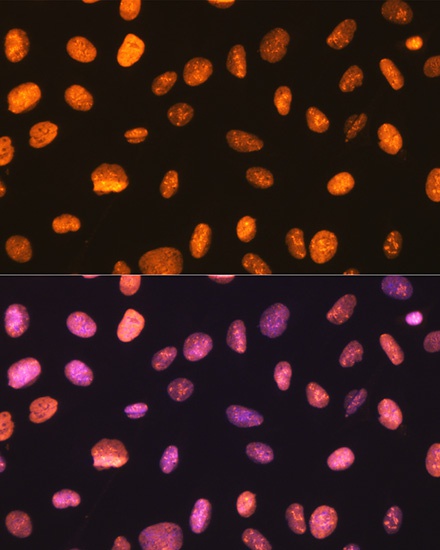
Immunofluorescence analysis of C6 cells using NONO/p54nrb Rabbit mAb (CAB3800) at dilution of 1:100 (40x lens). Secondary antibody: Cy3-conjugated Goat anti-Rabbit IgG (H+L) (AS007) at 1:500 dilution. Blue: DAPI for nuclear staining.
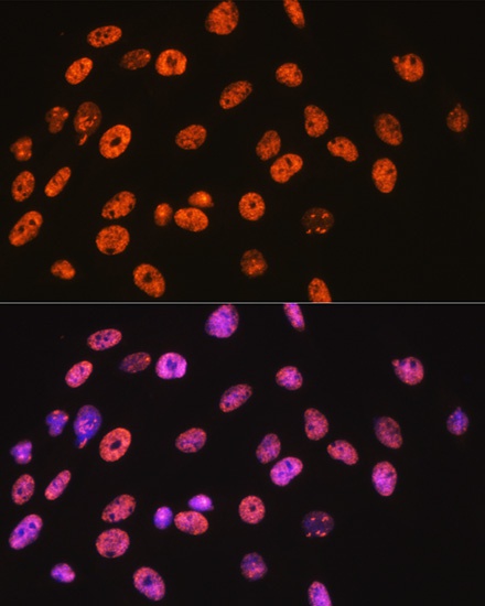
Immunofluorescence analysis of U-2 OS cells using NONO/p54nrb Rabbit mAb (CAB3800) at dilution of 1:100 (40x lens). Secondary antibody: Cy3-conjugated Goat anti-Rabbit IgG (H+L) (AS007) at 1:500 dilution. Blue: DAPI for nuclear staining.