The ENO2 Monoclonal Antibody (CAB19091) is a high-quality antibody developed for reliable detection and analysis of target proteins. This antibody, developed using rabbit monoclonal technology, is highly specific and sensitive for detecting NSE in human samples.NSE, also known as neuron-specific enolase, is a glycolytic enzyme found in neurons and neuroendocrine cells. It is often used as a biomarker for neuronal damage and tumor detection. The NSE Rabbit Monoclonal Antibody binds specifically to NSE protein, allowing for accurate detection and analysis in various experimental settings.
This antibody is validated for use in WB, IHC-P, IF/ICC, ELISA applications and has demonstrated reactivity against Human, Mouse, Rat samples.
Product Name:
ENO2 Monoclonal Antibody
SKU:
CAB19091
Size:
20μL, 100μL
Reactivity:
Human, Mouse, Rat
Clone Number:
ARC52246
Conjugate:
Unconjugated
Immunogen:
Recombinant protein (or fragment).This information is considered to be commercially sensitive.
Recommended starting concentration is 1 μg/mL. Please optimize the concentration based on your specific assay requirements.
Synonyms:
NSE, HEL-S-279, ENO2
Positive Sample:
SH-SY5Y, Mouse brain, Rat brain
Cellular Localization:
Cell Membrane, Cytoplasm.
Calculated MW:
47kDa
Observed MW:
47kDa
This gene encodes one of the three enolase isoenzymes found in mammals. This isoenzyme, a homodimer, is found in mature neurons and cells of neuronal origin. A switch from alpha enolase to gamma enolase occurs in neural tissue during development in rats and primates.
Purification Method
Affinity purification
Gene ID
2026
RRID
AB_2862583
Buffer Information
Store at -20℃. Avoid freeze / thaw cycles. Buffer: PBS containing 50% glycerol and 0.05% BSA, preserved with proclin300 or sodium azide, pH 7.3.
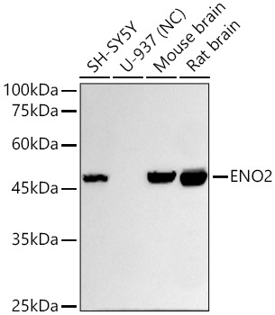
Western blot analysis of various lysates using ENO2 Rabbit mAb (CAB19091)at 1:10000 dilution incubated overnight at 4℃. Secondary antibody: HRP-conjugated Goat anti-Rabbit IgG (H+L) (CABS014) at 1:10000 dilution. Lysates/proteins: 25 μg per lane. Blocking buffer: 3% nonfat dry milk in TBST. Detection: ECL Basic Kit (AbGn00020). Negative control (NC): U-937 Exposure time: 15s.
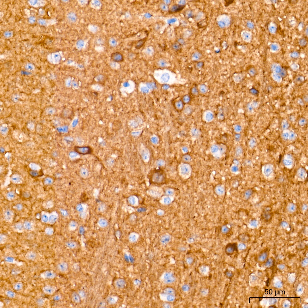
Immunohistochemistry analysis of paraffin-embedded Mouse brain tissue using ENO2 Rabbit mAb (CAB19091) at a dilution of 1:2000 (40x lens). High pressure antigen retrieval performed with 0.01M Tris-EDTA Buffer (pH 9.0) prior to IHC staining.
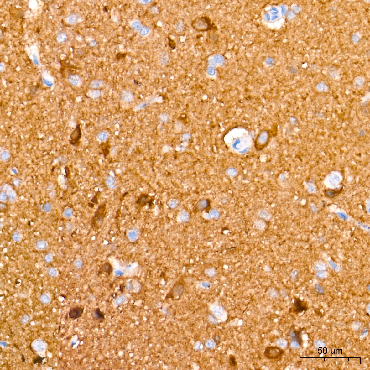
Immunohistochemistry analysis of paraffin-embedded Rat brain tissue using ENO2 Rabbit mAb (CAB19091) at a dilution of 1:2000 (40x lens). High pressure antigen retrieval performed with 0.01M Tris-EDTA Buffer (pH 9.0) prior to IHC staining.
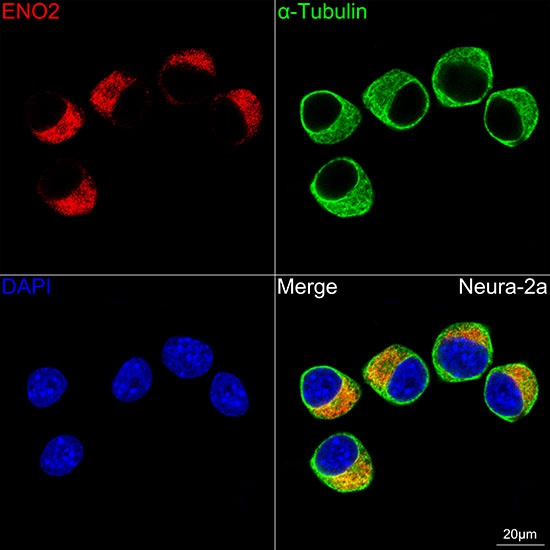
Confocal imaging of Neuro-2a cells using ENO2 Rabbit mAb (CAB19091,dilution 1:100)(Red). The cells were counterstained with α-Tubulin Mouse mAb (AC012,dilution 1:400) (Green). DAPI was used for nuclear staining (blue). Objective: 100x.