The NUMA1 Antibody (CAB0527) is a high-quality antibody developed for reliable detection and analysis of target proteins. This antibody, generated in rabbits, shows high reactivity towards human samples and is validated for use in Western blot applications. By binding specifically to the NUMA1 protein, this antibody allows for the detection and analysis of NUMA1 expression in various cell types, making it ideal for studies in cell biology and cancer research.NUMA1 is a key player in spindle organization and chromosome segregation during cell division, making it essential for maintaining genomic stability.
This antibody is validated for use in WB, IHC-P, IF/ICC, ELISA applications and has demonstrated reactivity against Human, Mouse, Rat samples.
Product Name:
NUMA1 Antibody
SKU:
CAB0527
Size:
20μL, 100μL
Reactivity:
Human, Mouse, Rat
Conjugate:
Unconjugated
Immunogen:
Recombinant protein (or fragment).This information is considered to be commercially sensitive.
Recommended starting concentration is 1 μg/mL. Please optimize the concentration based on your specific assay requirements.
Synonyms:
NUMA, NMP-22, NUMA1
Positive Sample:
HeLa, Mouse kidney
Cellular Localization:
Chromosome, Cytoplasm, Nucleus Matrix, Cytoskeleton, Spindle Pole.
Calculated MW:
238kDa
Observed MW:
238kDa
This gene encodes a large protein that forms a structural component of the nuclear matrix. The encoded protein interacts with microtubules and plays a role in the formation and organization of the mitotic spindle during cell division. Chromosomal translocation of this gene with the RARA (retinoic acid receptor, alpha) gene on chromosome 17 have been detected in patients with acute promyelocytic leukemia. Alternative splicing results in multiple transcript variants.
Purification Method
Affinity purification
Gene ID
4926
RRID
AB_2757245
Buffer Information
Store at -20℃. Avoid freeze / thaw cycles. Buffer: PBS containing 50% glycerol, preserved with proclin300 or sodium azide, pH 7.3.
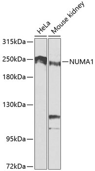
Western blot analysis of various lysates using NUMA1 Rabbit pAb (CAB0527) at 1:1000 dilution. Secondary antibody: HRP-conjugated Goat anti-Rabbit IgG (H+L) (CABS014) at 1:10000 dilution. Lysates/proteins: 25μg per lane. Blocking buffer: 3% nonfat dry milk in TBST. Detection: ECL Basic Kit (AbGn00020). Exposure time: 90s.
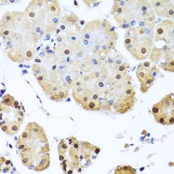
Immunohistochemistry analysis of paraffin-embedded Human stomach using NUMA1 Rabbit pAb (CAB0527) at dilution of 1:100 (40x lens). Microwave antigen retrieval performed with 0.01M PBS Buffer (pH 7.2) prior to IHC staining.
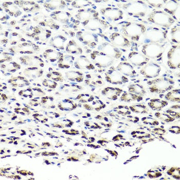
Immunohistochemistry analysis of paraffin-embedded Mouse stomach using NUMA1 Rabbit pAb (CAB0527) at dilution of 1:100 (40x lens). Microwave antigen retrieval performed with 0.01M PBS Buffer (pH 7.2) prior to IHC staining.
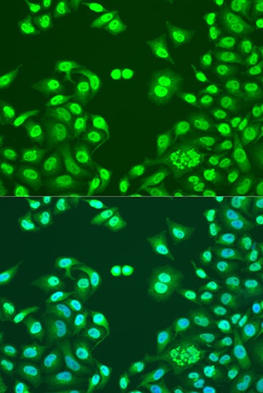
Immunofluorescence analysis of U2OS cells using NUMA1 Rabbit pAb (CAB0527) at dilution of 1:100. Secondary antibody: Cy3-conjugated Goat anti-Rabbit IgG (H+L) (CABS007) at 1:500 dilution. Blue: DAPI for nuclear staining.