The OAT Antibody (CAB6235) is a high-quality antibody developed for reliable detection and analysis of target proteins. This antibody, produced in rabbits, is highly specific to human OAT and is suitable for use in Western blot applications. By targeting the OAT protein, this antibody allows for the detection and analysis of OAT levels in various cell types, making it a useful tool for studies in biochemistry and metabolism.OAT is an essential enzyme that plays a crucial role in the urea cycle, which is responsible for the detoxification of ammonia in the body.
This antibody is validated for use in WB, IHC-P, IP, ELISA applications and has demonstrated reactivity against Human, Mouse, Rat samples.
Product Name:
OAT Antibody
SKU:
CAB6235
Size:
20μL, 100μL
Reactivity:
Human, Mouse, Rat
Conjugate:
Unconjugated
Immunogen:
Recombinant protein (or fragment).This information is considered to be commercially sensitive.
0.5μg-4μg antibody for 200μg-400μg extracts of whole cells
ELISA
Recommended starting concentration is 1 μg/mL. Please optimize the concentration based on your specific assay requirements.
Synonyms:
OKT, GACR, HOGA, OATASE, OAT
Positive Sample:
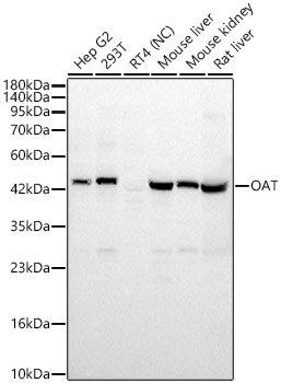
Hep G2, 293T, Mouse liver, Mouse kidney, Rat liver
Cellular Localization:
Mitochondrion Matrix.
Calculated MW:
49kDa
Observed MW:
49kDa
This gene encodes the mitochondrial enzyme ornithine aminotransferase, which is a key enzyme in the pathway that converts arginine and ornithine into the major excitatory and inhibitory neurotransmitters glutamate and GABA. Mutations that result in a deficiency of this enzyme cause the autosomal recessive eye disease Gyrate Atrophy. Alternatively spliced transcript variants encoding different isoforms have been described. Related pseudogenes have been defined on the X chromosome.
Purification Method
Affinity purification
Gene ID
4942
RRID
AB_2766843
Buffer Information
Store at -20℃. Avoid freeze / thaw cycles. Buffer: PBS containing 50% glycerol, preserved with proclin300 or sodium azide, pH 7.3.
Western blot analysis of various lysates using OAT Rabbit pAb (CAB6235) at 1:2000 dilution. Secondary antibody: HRP-conjugated Goat anti-Rabbit IgG (H+L) (CABS014) at 1:10000 dilution. Lysates/proteins: 25 μg per lane. Blocking buffer: 3% nonfat dry milk in TBST. Detection: ECL Basic Kit (AbGn00020). Negative control (NC): RT4. Exposure time: 30s.
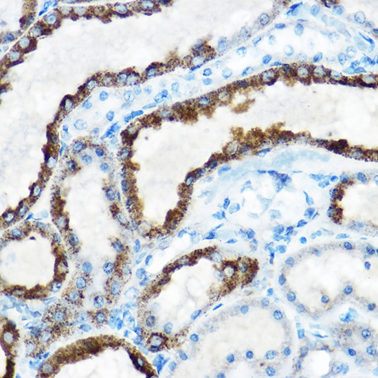
Immunohistochemistry analysis of paraffin-embedded Rat kidney using OAT Rabbit pAb (CAB6235) at dilution of 1:100 (40x lens). High pressure antigen retrieval performed with 0.01M Citrate buffer (pH 6.0) prior to IHC staining.
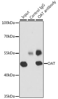
Immunoprecipitation analysis of 200 μg extracts of MCF-7 cells, using 3 μg OAT antibody (CAB6235). Western blot was performed from the immunoprecipitate using OAT antibody (CAB6235) at a dilution of 1:1000.