Pan Phospho-Tyrosine Monoclonal Antibody (CABP1162)
The Pan Phospho-Tyrosine Monoclonal Antibody (CABP1162) is a high-quality antibody developed for reliable detection and analysis of target proteins. This antibody specifically recognizes phosphorylated tyrosine residues on a wide range of proteins, allowing for the detection and analysis of tyrosine phosphorylation in various cell types.Raised in rabbits, this monoclonal antibody is highly sensitive and specific for human samples, making it an ideal choice for Western blot applications. By targeting phosphorylated tyrosine residues, researchers can gain insights into the activation of key signaling molecules involved in processes such as cell growth, differentiation, and apoptosis.
This antibody is validated for use in WB, IP, ELISA applications and has demonstrated reactivity against Human, Mouse, Rat, Other (Wide Range Predicted) samples.
Product Name:
Pan Phospho-Tyrosine Monoclonal Antibody
SKU:
CABP1162
Size:
20μL, 100μL
Reactivity:
Human, Mouse, Rat, Other (Wide Range Predicted)
Clone Number:
ARC54383
Conjugate:
Unconjugated
Immunogen:
Synthetic peptide. This information is considered to be commercially sensitive.
Sequence:
Email for sequence
Tested Applications:
WBIPELISA
Recommended Dilution:
WB
1:500 - 1:1000
IP
0.5μg-4μg antibody for 200μg-400μg extracts of whole cells
ELISA
Recommended starting concentration is 1 μg/mL. Please optimize the concentration based on your specific assay requirements.
Positive Sample:
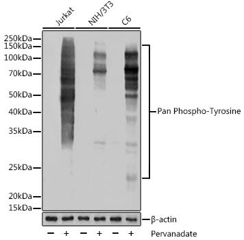
Jurkat treated with Pervanadate, NIH/3T3 treated with Pervanadate, C6 treated with Pervanadate,
Tyrosine phosphorylation (pTyr), much of which occurred on localized multiple sites, initiates cellular signaling, governs cellular functions, and its dysregulation is implicated in many diseases, especially cancers. pTyr-specific sensing is of great significance for understanding disease states and developing targeted anticancer drugs.
Purification Method
Affinity purification
RRID
AB_2864021
Buffer Information
Store at -20℃. Avoid freeze / thaw cycles. Buffer: PBS with 0.09% sodium azide,0.05% BSA,50% glycerol,pH7.3.
Western blot analysis of various lysates using Pan Phospho-Tyrosine Rabbit mAb (CABP1162) at 1:1000 dilution. Jurkat cells and NIH/3T3 cells and C6 cells were treated with Pervanadate (1 mM) at 37℃ for 30 minutes. Secondary antibody: HRP-conjugated Goat anti-Rabbit IgG (H+L) (CABS014) at 1:10000 dilution. Lysates/proteins: 25μg per lane. Blocking buffer: 3% BSA. Detection: ECL Basic Kit (AbGn00020). Exposure time: 5s.
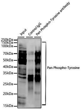
Immunoprecipitation analysis of 300 μg extracts of Jurkat (pervanadate) cells using 3 μg Pan Phospho-Tyrosine Rabbit mAb (CABP1162). Western blot was performed from the immunoprecipitate using Pan Phospho-Tyrosine Rabbit mAb (CABP1162) at a dilution of 1:1000.