The PAX3 Monoclonal Antibody (CAB22293) is a high-quality antibody developed for reliable detection and analysis of target proteins. This highly specific antibody, produced using hybridoma technology, is reactive with human samples and is validated for use in various applications, including immunohistochemistry and flow cytometry.Pax3 is a key regulator of embryonic development, specifically in the formation of skeletal muscles and neural crest cells. Aberrant expression or function of Pax3 has been implicated in various muscular dystrophies and cancers, making it a valuable target for studies in developmental biology and oncology.
This antibody is validated for use in WB, ELISA applications and has demonstrated reactivity against Human, Mouse, Rat samples.
Product Name:
PAX3 Monoclonal Antibody
SKU:
CAB22293
Size:
20μL, 100μL
Reactivity:
Human, Mouse, Rat
Clone Number:
ARC57259
Conjugate:
Unconjugated
Immunogen:
Synthetic peptide. This information is considered to be commercially sensitive.
Recommended starting concentration is 1 μg/mL. Please optimize the concentration based on your specific assay requirements.
Synonyms:
WS1, WS3, CDHS, HUP2, PAX-3, PAX3
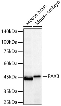
Positive Sample:
Mouse brain, Mouse embryo
Cellular Localization:
Nucleus.
Calculated MW:
53kDa
Observed MW:
53kDa
This gene is a member of the paired box (PAX) family of transcription factors. Members of the PAX family typically contain a paired box domain and a paired-type homeodomain. These genes play critical roles during fetal development. Mutations in paired box gene 3 are associated with Waardenburg syndrome, craniofacial-deafness-hand syndrome, and alveolar rhabdomyosarcoma. The translocation t(2;13)(q35;q14), which represents a fusion between PAX3 and the forkhead gene, is a frequent finding in alveolar rhabdomyosarcoma. Alternative splicing results in transcripts encoding isoforms with different C-termini.
Purification Method
Affinity purification
Gene ID
5077
Buffer Information
Store at -20℃. Avoid freeze / thaw cycles. Buffer: PBS containing 50% glycerol and 0.05% BSA, preserved with proclin300 or sodium azide, pH 7.3.
Western blot analysis of various lysates using PAX3 Rabbit mAb (CAB22293) at 1:1000 dilution incubated overnight at 4℃. Secondary antibody: HRP-conjugated Goat anti-Rabbit IgG (H+L) (CABS014) at 1:10000 dilution. Lysates/proteins: 25 μg per lane. Blocking buffer: 3% nonfat dry milk in TBST. Detection: ECL Basic Kit (AbGn00020). Exposure time: 30s.
 at 1:600 dilution. Secondary antibody: HRP Goat Anti-Rabbit IgG (H+L) at 1:10000 dilution. Lysates/proteins: 25μg per lane. Blocking buffer: 3% nonfat dry milk in TBST.")
 at 1:600 dilution. Secondary antibody: HRP Goat Anti-Rabbit IgG (H+L) at 1:10000 dilution. Lysates/proteins: 25μg per lane. Blocking buffer: 3% nonfat dry milk in TBST.")


")