The PAX3 Antibody (CAB13930) is a high-quality antibody developed for reliable detection and analysis of target proteins. This antibody, generated in rabbits, exhibits high specificity and sensitivity towards human samples, making it a reliable tool for Western blot applications. By binding to the PAX3 protein, this antibody enables precise detection and analysis in a variety of cell types, making it an essential resource for researchers in fields such as developmental biology and cancer research.PAX3 is known for its role in regulating the development of tissues and organs, particularly in the formation of skeletal muscles and the nervous system.
This antibody is validated for use in WB, ELISA applications and has demonstrated reactivity against Human, Mouse, Rat samples.
Product Name:
PAX3 Antibody
SKU:
CAB13930
Size:
20μL, 100μL
Reactivity:
Human, Mouse, Rat
Conjugate:
Unconjugated
Immunogen:
Recombinant protein (or fragment).This information is considered to be commercially sensitive.
Recommended starting concentration is 1 μg/mL. Please optimize the concentration based on your specific assay requirements.
Synonyms:
WS1, WS3, CDHS, HUP2, PAX-3, PAX3
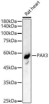
Positive Sample:
Rat heart
Cellular Localization:
Nucleus.
Calculated MW:
53kDa
Observed MW:
53kDa
This gene is a member of the paired box (PAX) family of transcription factors. Members of the PAX family typically contain a paired box domain and a paired-type homeodomain. These genes play critical roles during fetal development. Mutations in paired box gene 3 are associated with Waardenburg syndrome, craniofacial-deafness-hand syndrome, and alveolar rhabdomyosarcoma. The translocation t(2;13)(q35;q14), which represents a fusion between PAX3 and the forkhead gene, is a frequent finding in alveolar rhabdomyosarcoma. Alternative splicing results in transcripts encoding isoforms with different C-termini.
Purification Method
Affinity purification
Gene ID
5077
RRID
AB_2760782
Buffer Information
Store at -20℃. Avoid freeze / thaw cycles. Buffer: PBS containing 50% glycerol, preserved with proclin300 or sodium azide, pH 7.3.
Western blot analysis of lysates from Rat heart, using PAX3 Rabbit pAb (CAB13930) at 1:500 dilution. Secondary antibody: HRP-conjugated Goat anti-Rabbit IgG (H+L) (CABS014) at 1:10000 dilution. Lysates/proteins: 25μg per lane. Blocking buffer: 3% nonfat dry milk in TBST. Detection: ECL Basic Kit (AbGn00020). Exposure time: 90s.