The PAX3 Antibody (CAB1675) is a high-quality antibody developed for reliable detection and analysis of target proteins. This antibody is raised in rabbits and has been validated for use in Western blot applications, providing reliable detection and analysis of Pax3 in various cell types.Pax3 is known for its role in neural crest development, muscle development, and melanocyte differentiation, making it a crucial target for investigation in developmental biology and regenerative medicine research. By targeting Pax3 with this antibody, researchers can gain insights into its functions and pathways, ultimately leading to a better understanding of its role in tissue regeneration, disease progression, and potential therapeutic applications.
This antibody is validated for use in WB, IHC-P, IF/ICC, ELISA applications and has demonstrated reactivity against Human, Mouse, Rat samples.
Product Name:
PAX3 Antibody
SKU:
CAB1675
Size:
20μL, 100μL
Reactivity:
Human, Mouse, Rat
Conjugate:
Unconjugated
Immunogen:
Recombinant protein (or fragment).This information is considered to be commercially sensitive.
Recommended starting concentration is 1 μg/mL. Please optimize the concentration based on your specific assay requirements.
Synonyms:
WS1, WS3, CDHS, HUP2, PAX-3, PAX3
Positive Sample:
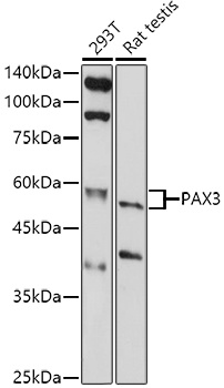
293T, Rat testis
Cellular Localization:
Nucleus.
Calculated MW:
53kDa
Observed MW:
60kDa
This gene is a member of the paired box (PAX) family of transcription factors. Members of the PAX family typically contain a paired box domain and a paired-type homeodomain. These genes play critical roles during fetal development. Mutations in paired box gene 3 are associated with Waardenburg syndrome, craniofacial-deafness-hand syndrome, and alveolar rhabdomyosarcoma. The translocation t(2;13)(q35;q14), which represents a fusion between PAX3 and the forkhead gene, is a frequent finding in alveolar rhabdomyosarcoma. Alternative splicing results in transcripts encoding isoforms with different C-termini.
Purification Method
Affinity purification
Gene ID
5077
RRID
AB_2763730
Buffer Information
Store at -20℃. Avoid freeze / thaw cycles. Buffer: PBS containing 50% glycerol, preserved with proclin300 or sodium azide, pH 7.3.
Western blot analysis of various lysates using PAX3 Rabbit pAb (CAB1675) at 1:1000 dilution. Secondary antibody: HRP-conjugated Goat anti-Rabbit IgG (H+L) (CABS014) at 1:10000 dilution. Lysates/proteins: 25μg per lane. Blocking buffer: 3% nonfat dry milk in TBST. Detection: ECL Basic Kit (AbGn00020). Exposure time: 180s.
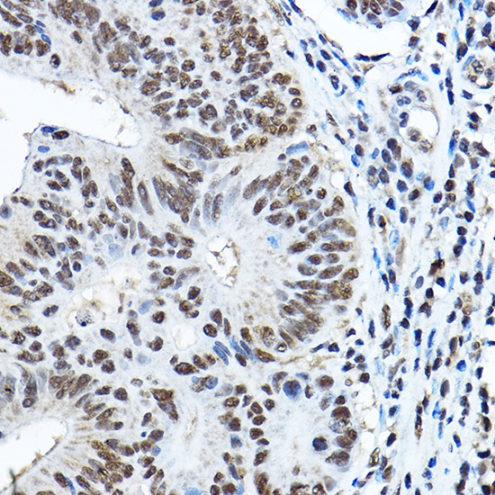
Immunohistochemistry analysis of paraffin-embedded Human colon carcinoma using PAX3 Rabbit pAb (CAB1675) at dilution of 1:100 (40x lens). High pressure antigen retrieval performed with 0.01M Citrate buffer (pH 6.0) prior to IHC staining.