The PCNA Monoclonal Antibody (CAB12427) is a high-quality antibody developed for reliable detection and analysis of target proteins. This antibody, developed using rabbit monoclonal technology, is highly specific and sensitive for detecting PCNA in a variety of samples, making it suitable for applications such as immunohistochemistry and immunofluorescence.PCNA is a marker of cell proliferation and is often upregulated in cancer cells, making it a target of interest for cancer research. By using the PCNA Rabbit Monoclonal Antibody, researchers can easily visualize and quantify PCNA levels in cells and tissues, providing valuable insights into cell cycle progression and DNA repair mechanisms.
This antibody is validated for use in WB, IHC-P, ELISA applications and has demonstrated reactivity against Human, Mouse, Rat, African green monkey samples.
Product Name:
PCNA Monoclonal Antibody
SKU:
CAB12427
Size:
20μL, 100μL
Reactivity:
Human, Mouse, Rat, African green monkey
Clone Number:
ARC51324
Conjugate:
Unconjugated
Immunogen:
Synthetic peptide. This information is considered to be commercially sensitive.
Recommended starting concentration is 1 μg/mL. Please optimize the concentration based on your specific assay requirements.
Synonyms:
ATLD2, PCNA
Positive Sample:
HeLa, 293T, MCF7, C2C12, C6
Cellular Localization:
Nucleus.
Calculated MW:
29kDa
Observed MW:
36kDa
The protein encoded by this gene is found in the nucleus and is a cofactor of DNA polymerase delta. The encoded protein acts as a homotrimer and helps increase the processivity of leading strand synthesis during DNA replication. In response to DNA damage, this protein is ubiquitinated and is involved in the RAD6-dependent DNA repair pathway. Two transcript variants encoding the same protein have been found for this gene. Pseudogenes of this gene have been described on chromosome 4 and on the X chromosome.
Purification Method
Affinity purification
Gene ID
5111
RRID
AB_2861664
Buffer Information
Store at -20℃. Avoid freeze / thaw cycles. Buffer: PBS with 0.09% Sodium azide,0.05% BSA,50% glycerol,pH7.3.
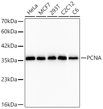
Western blot analysis of various lysates using PCNA Rabbit mAb (CAB12427) at 1:5000 dilution incubated overnight at 4℃. Secondary antibody: HRP-conjugated Goat anti-Rabbit IgG (H+L) (CABS014) at 1:10000 dilution. Lysates/proteins: 25 μg per lane. Blocking buffer: 3% nonfat dry milk in TBST. Detection: ECL Basic Kit (AbGn00020). Exposure time: 20s.
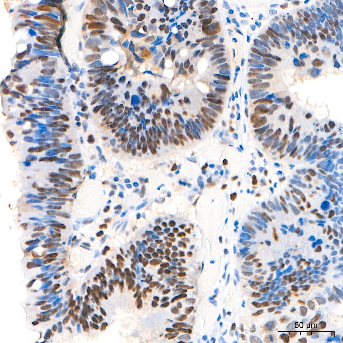
Immunohistochemistry analysis of paraffin-embedded Human colon carcinoma tissue using PCNA Rabbit mAb (CAB12427) at a dilution of 1:15000 (40x lens). High pressure antigen retrieval performed with 0.01M Tris-EDTA Buffer (pH 9.0) prior to IHC staining.
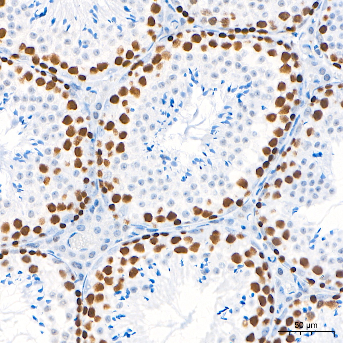
Immunohistochemistry analysis of paraffin-embedded Mouse testis tissue using PCNA Rabbit mAb (CAB12427) at a dilution of 1:15000 (40x lens). High pressure antigen retrieval performed with 0.01M Tris-EDTA Buffer (pH 9.0) prior to IHC staining.
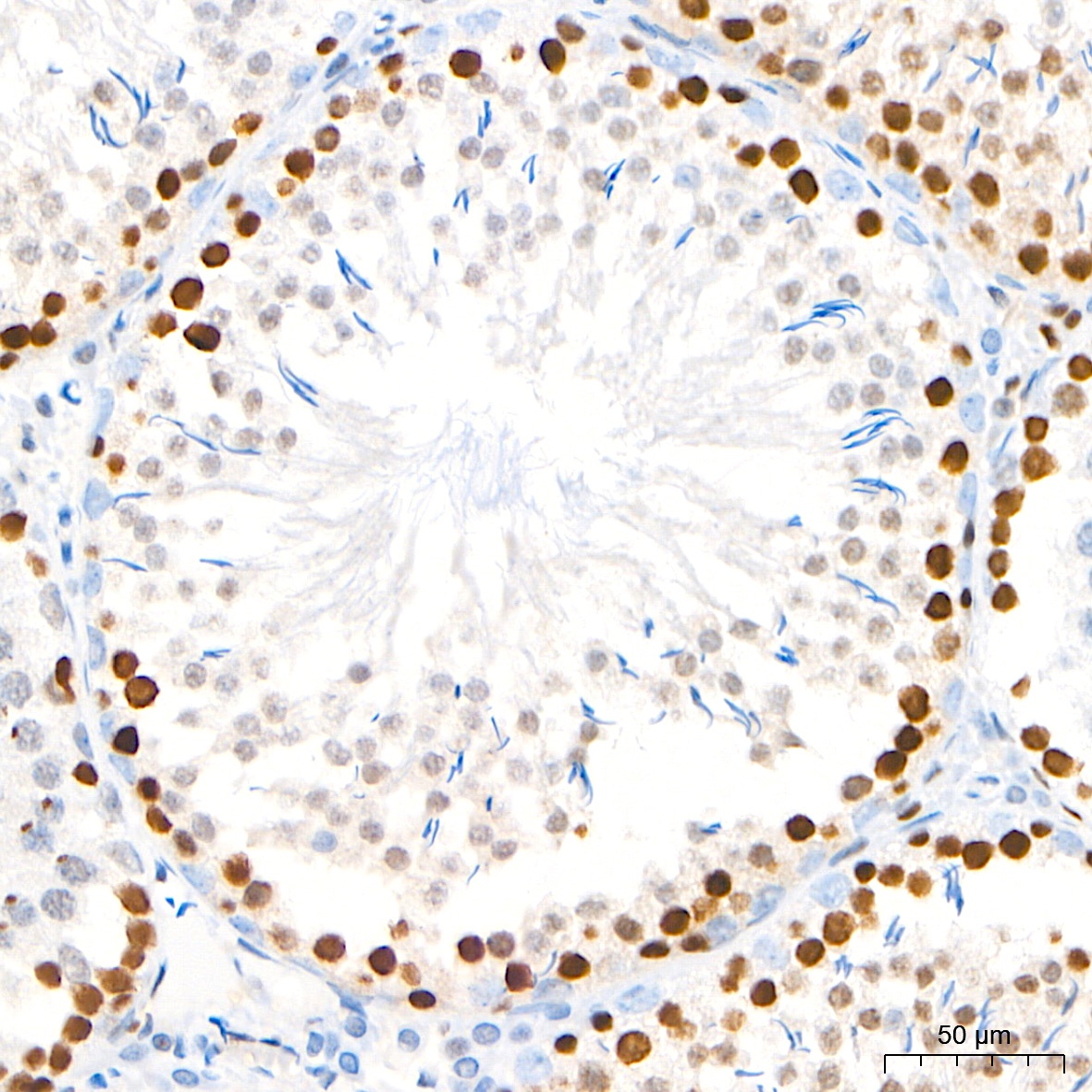
Immunohistochemistry analysis of paraffin-embedded Rat testis tissue using PCNA Rabbit mAb (CAB12427) at a dilution of 1:15000 (40x lens). High pressure antigen retrieval performed with 0.01M Tris-EDTA Buffer (pH 9.0) prior to IHC staining.


")
")


