The PDE10A Antibody (CAB15597) is a high-quality antibody developed for reliable detection and analysis of target proteins. This antibody, produced in rabbits, exhibits high reactivity with human samples and is validated for use in Western blot applications.PDE10A is involved in various cellular processes, including signal transduction and neurotransmission, making it a target of interest in studies related to neurological disorders and psychiatric conditions. By binding specifically to the PDE10A protein, this antibody enables researchers to detect and analyze its expression in different cell types, providing insights into its function and potential therapeutic targeting.
This antibody is validated for use in WB, IF/ICC, ELISA applications and has demonstrated reactivity against Human, Rat samples.
Product Name:
PDE10A Antibody
SKU:
CAB15597
Size:
20μL, 100μL
Reactivity:
Human, Rat
Conjugate:
Unconjugated
Immunogen:
Recombinant protein (or fragment).This information is considered to be commercially sensitive.
The protein encoded by this gene belongs to the cyclic nucleotide phosphodiesterase family. It plays a role in signal transduction by regulating the intracellular concentration of cyclic nucleotides. This protein can hydrolyze both cAMP and cGMP to the corresponding nucleoside 5' monophosphate, but has higher affinity for cAMP, and is more efficient with cAMP as substrate. Alternatively spliced transcript variants have been described for this gene.
Purification Method
Affinity purification
Gene ID
10846
RRID
AB_2763002
Buffer Information
Store at -20℃. Avoid freeze / thaw cycles. Buffer: Buffer: PBS containing 50% glycerol, preserved with proclin300 or sodium azide, pH 7.3.
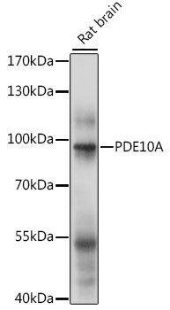
Western blot analysis of lysates from rat brain, using PDE10A Rabbit pAb (CAB15597) at 1:1000 dilution. Secondary antibody: HRP-conjugated Goat anti-Rabbit IgG (H+L) (CABS014) at 1:10000 dilution. Lysates/proteins: 25μg per lane. Blocking buffer: 3% nonfat dry milk in TBST. Detection: ECL Basic Kit (AbGn00020). Exposure time: 5s.
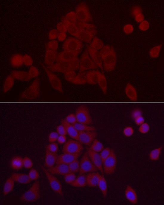
Immunofluorescence analysis of HeLa cells using PDE10A Rabbit pAb (CAB15597) at dilution of 1:20 (40x lens). Secondary antibody: Cy3-conjugated Goat anti-Rabbit IgG (H+L) (CABS007) at 1:500 dilution. Blue: DAPI for nuclear staining.