The [KO Validated] PDPK1 Antibody (CAB1665) is a high-quality antibody developed for reliable detection and analysis of target proteins. Enables 3-phosphoinositide-dependent protein kinase activity; phospholipase activator activity; and phospholipase binding activity. Involved in several processes, including cell surface receptor signaling pathway; regulation of protein kinase activity; and regulation of signal transduction. Acts upstream of or within intracellular signal transduction. Located in cell projection; cytosol; and plasma membrane. Implicated in prostate cancer. Biomarker of lung non-small cell carcinoma. RRID AB_2763720 Gene ID 5170 Swiss Prot Synonym PDK1; PDPK2; PDPK2P; PRO0461; K1
This antibody is validated for use in WB, IHC-P, ELISA applications and has demonstrated reactivity against Human, Mouse, Rat samples.
Product Name:
[KO Validated] PDPK1 Antibody
SKU:
CAB1665
Size:
100μL, 20μL
Reactivity:
Human, Mouse, Rat
Clone Number:
-
Conjugate:
Unconjugated
Immunogen:
Recombinant protein (or fragment).This information is considered to be commercially sensitive.
Tested Applications:
WBIHC-PELISA
Recommended Dilution:
WB
1:500 - 1:1000
IHC-P
1:50 - 1:200
ELISA
Recommended starting concentration is 1 μg/mL. Please optimize the concentration based on your specific assay requirements.
Enables 3-phosphoinositide-dependent protein kinase activity; phospholipase activator activity; and phospholipase binding activity. Involved in several processes, including cell surface receptor signaling pathway; regulation of protein kinase activity; and regulation of signal transduction. Acts upstream of or within intracellular signal transduction. Located in cell projection; cytosol; and plasma membrane. Implicated in prostate cancer. Biomarker of lung non-small cell carcinoma. RRID AB_2763720 Gene ID 5170 Swiss Prot Synonym PDK1; PDPK2; PDPK2P; PRO0461; K1
Purification Method:
Affinity purification
Gene ID:
5170
RRID:
AB_2763720
Buffer Information:
Store at -20℃. Avoid freeze / thaw cycles. Buffer: PBS containing 50% glycerol, preserved with proclin300 or sodium azide, pH 7.3.
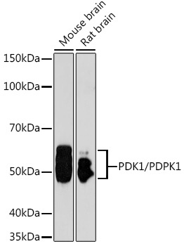
Western blot analysis of various lysates using [KO Validated] PDK1/PDPK1 Rabbit pAb (CAB1665) at 1:1000 dilution. Secondary antibody: HRP-conjugated Goat anti-Rabbit IgG (H+L) (AS014) at 1:10000 dilution. Lysates/proteins: 25μg per lane. Blocking buffer: 3% nonfat dry milk in TBST. Detection: ECL Basic Kit (AbGn00020). Exposure time: 90s.
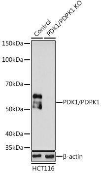
Western blot analysis of lysates from wild type (WT) and PDK1/PDPK1 knockout (KO) HCT116 cells, using [KO Validated] PDK1/PDPK1 Rabbit pAb (CAB1665) at 1:1000 dilution. Secondary antibody: HRP-conjugated Goat anti-Rabbit IgG (H+L) (AS014) at 1:10000 dilution. Lysates/proteins: 25μg per lane. Blocking buffer: 3% nonfat dry milk in TBST. Detection: ECL Basic Kit (AbGn00020). Exposure time: 90s.