The PEG10 Antibody (CAB2787) is a high-quality antibody developed for reliable detection and analysis of target proteins. This is a paternally expressed imprinted gene that is thought to have been derived from the Ty3/Gypsy family of retrotransposons. It contains two overlapping open reading frames, RF1 and RF2, and expresses two proteins: a shorter, gag-like protein (with a CCHC-type zinc finger domain) from RF1; and a longer, gag/pol-like fusion protein (with an additional aspartic protease motif) from RF1/RF2 by -1 translational frameshifting (-1 FS). While -1 FS has been observed in RNA viruses and transposons in both prokaryotes and eukaryotes, this gene represents the first example of -1 FS in a eukaryotic cellular gene. This gene is highly conserved across mammalian species and retains the heptanucleotide (GGGAAAC) and pseudoknot elements required for -1 FS. It is expressed in adult and embryonic tissues (most notably in placenta) and reported to have a role in cell proliferation, differentiation and apoptosis. Overexpression of this gene has been associated with several malignancies, such as hepatocellular carcinoma and B-cell lymphocytic leukemia. Knockout mice lacking this gene showed early embryonic lethality with placental defects, indicating the importance of this gene in embryonic development. Additional isoforms resulting from alternatively spliced transcript variants, and use of upstream non-AUG (CUG) start codon have been reported for this gene.
This antibody is validated for use in WB, IHC-P, ELISA applications and has demonstrated reactivity against Human, Mouse, Rat samples.
Product Name:
PEG10 Antibody
SKU:
CAB2787
Size:
100μL, 20μL
Reactivity:
Human, Mouse, Rat
Conjugate:
Unconjugated
Immunogen:
Recombinant protein (or fragment).This information is considered to be commercially sensitive.
Tested Applications:
WBIHC-PELISA
Recommended Dilution:
WB
1:200 - 1:2000
IHC-P
1:50 - 1:200
ELISA
Recommended starting concentration is 1 μg/mL. Please optimize the concentration based on your specific assay requirements.
This is a paternally expressed imprinted gene that is thought to have been derived from the Ty3/Gypsy family of retrotransposons. It contains two overlapping open reading frames, RF1 and RF2, and expresses two proteins: a shorter, gag-like protein (with a CCHC-type zinc finger domain) from RF1; and a longer, gag/pol-like fusion protein (with an additional aspartic protease motif) from RF1/RF2 by -1 translational frameshifting (-1 FS). While -1 FS has been observed in RNA viruses and transposons in both prokaryotes and eukaryotes, this gene represents the first example of -1 FS in a eukaryotic cellular gene. This gene is highly conserved across mammalian species and retains the heptanucleotide (GGGAAAC) and pseudoknot elements required for -1 FS. It is expressed in adult and embryonic tissues (most notably in placenta) and reported to have a role in cell proliferation, differentiation and apoptosis. Overexpression of this gene has been associated with several malignancies, such as hepatocellular carcinoma and B-cell lymphocytic leukemia. Knockout mice lacking this gene showed early embryonic lethality with placental defects, indicating the importance of this gene in embryonic development. Additional isoforms resulting from alternatively spliced transcript variants, and use of upstream non-AUG (CUG) start codon have been reported for this gene.
Purification Method
Affinity purification
Gene ID
23089
RRID
AB_2764629
Buffer Information
Store at -20℃. Avoid freeze / thaw cycles. Buffer: PBS containing 50% glycerol, preserved with proclin300 or sodium azide, pH 7.3.
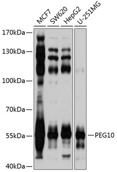
Western blot analysis of various lysates using PEG10 Rabbit pAb (CAB2787) at 1:1000 dilution. Secondary antibody: HRP-conjugated Goat anti-Rabbit IgG (H+L) (AS014) at 1:10000 dilution. Lysates/proteins: 25μg per lane. Blocking buffer: 3% nonfat dry milk in TBST. Detection: ECL Basic Kit (AbGn00020). Exposure time: 1s.
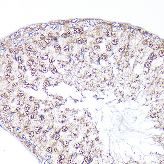
Immunohistochemistry analysis of paraffin-embedded Rat testis using PEG10 Rabbit pAb (CAB2787) at dilution of 1:100 (40x lens). High pressure antigen retrieval performed with 0.01M Citrate buffer (pH 6.0) prior to IHC staining.
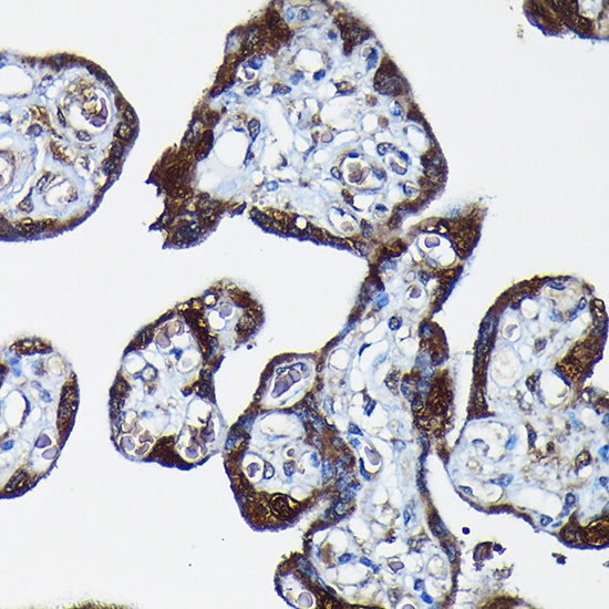
Immunohistochemistry analysis of paraffin-embedded Human placenta using PEG10 Rabbit pAb (CAB2787) at dilution of 1:100 (40x lens). High pressure antigen retrieval performed with 0.01M Citrate buffer (pH 6.0) prior to IHC staining.
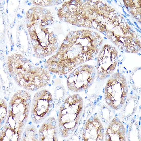
Immunohistochemistry analysis of paraffin-embedded Mouse kidney using PEG10 Rabbit pAb (CAB2787) at dilution of 1:100 (40x lens). High pressure antigen retrieval performed with 0.01M Citrate buffer (pH 6.0) prior to IHC staining.