The PGD Monoclonal Antibody (CAB0563) is a high-quality antibody developed for reliable detection and analysis of target proteins. This antibody, produced in rabbits, exhibits high reactivity with human samples and is validated for use in Western blot applications. By specifically binding to the PGD protein, this antibody allows for the detection and analysis of PGD expression in various cell types.PGD, also known as prostaglandin D2 synthase, plays a key role in the production of prostaglandins, lipid compounds that function as signaling molecules in inflammation and immune responses.
This antibody is validated for use in WB, IF/ICC, IP, ELISA applications and has demonstrated reactivity against Human, Mouse, Rat samples.
Product Name:
PGD Monoclonal Antibody
SKU:
CAB0563
Size:
20μL, 100μL
Reactivity:
Human, Mouse, Rat
Clone Number:
ARC2516
Conjugate:
Unconjugated
Immunogen:
Synthetic peptide. This information is considered to be commercially sensitive.
Sequence:
PELQ NLLL DDFF KSAV ENCQ DSWR RAVS TGVQ AGIP MPCF TTAL SFYD GYRH EMLP ASLI QAQR DYFG AHTY ELLA KPGQ FIHT NWTG HGGT VSSS SYNA
Tested Applications:
WBIF/ICCIPELISA
Recommended Dilution:
WB
1:500 - 1:1000
IF/ICC
1:50 - 1:200
IP
0.5μg-4μg antibody for 200μg-400μg extracts of whole cells
ELISA
Recommended starting concentration is 1 μg/mL. Please optimize the concentration based on your specific assay requirements.
Synonyms:
6PGD, PGD
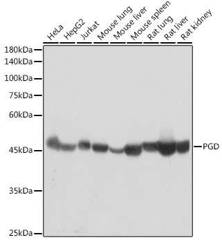
Positive Sample:
HeLa, HepG2, Jurkat, Mouse lung, Mouse liver, Mouse spleen, Rat lung, Rat liver, Rat kidney
Cellular Localization:
Cytoplasm.
Calculated MW:
53kDa
Observed MW:
49kDa
6-phosphogluconate dehydrogenase is the second dehydrogenase in the pentose phosphate shunt. Deficiency of this enzyme is generally asymptomatic, and the inheritance of this disorder is autosomal dominant. Hemolysis results from combined deficiency of 6-phosphogluconate dehydrogenase and 6-phosphogluconolactonase suggesting a synergism of the two enzymopathies. Several transcript variants encoding different isoforms have been found for this gene.
Purification Method
Affinity purification
Gene ID
5226
Buffer Information
Store at -20℃. Avoid freeze / thaw cycles. Buffer: PBS containing 50% glycerol and 0.05% BSA, preserved with proclin300 or sodium azide, pH 7.3.
Western blot analysis of various lysates using PGD Rabbit mAb (CAB0563) at 1:1000 dilution. Secondary antibody: HRP-conjugated Goat anti-Rabbit IgG (H+L) (CABS014) at 1:10000 dilution. Lysates/proteins: 25μg per lane. Blocking buffer: 3% nonfat dry milk in TBST. Detection: ECL Basic Kit (AbGn00020). Exposure time: 5s.
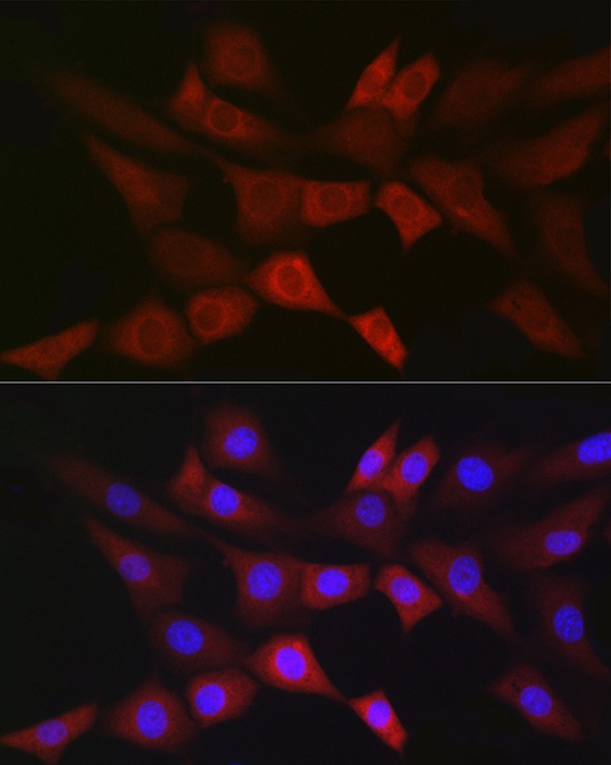
Immunofluorescence analysis of NIH/3T3 cells using PGD Rabbit mAb (CAB0563) at dilution of 1:100 (40x lens). Secondary antibody: Cy3-conjugated Goat anti-Rabbit IgG (H+L) (CABS007) at 1:500 dilution. Blue: DAPI for nuclear staining.
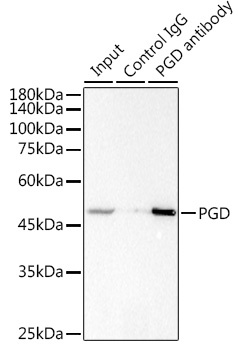
Immunoprecipitation analysis of 300 μg extracts of Jurkat cells using 3 μg PGD antibody (CAB0563). Western blot was performed from the immunoprecipitate using PGD antibody (CAB0563) at a dilution of 1:1000.