The PHGDH Antibody (CAB10461) is a high-quality antibody developed for reliable detection and analysis of target proteins. This antibody, produced in rabbits, is highly specific and reactive to human samples, making it ideal for applications such as Western blotting.PHGDH is an essential enzyme involved in cancer metabolism, as it is often upregulated in cancer cells to support their rapid growth. By targeting PHGDH with this antibody, researchers can gain insights into the role of this enzyme in cancer progression and potentially identify new therapeutic targets.
This antibody is validated for use in WB, IF/ICC, IP, ELISA applications and has demonstrated reactivity against Human, Mouse, Rat samples.
Product Name:
PHGDH Antibody
SKU:
CAB10461
Size:
20μL, 100μL
Reactivity:
Human, Mouse, Rat
Conjugate:
Unconjugated
Immunogen:
Recombinant protein (or fragment).This information is considered to be commercially sensitive.
This gene encodes the enzyme which is involved in the early steps of L-serine synthesis in animal cells. L-serine is required for D-serine and other amino acid synthesis. The enzyme requires NAD/NADH as a cofactor and forms homotetramers for activity. Mutations in this gene have been found in a family with congenital microcephaly, psychomotor retardation and other symptoms. Multiple alternatively spliced transcript variants have been found, however the full-length nature of most are not known.
Purification Method
Affinity purification
Gene ID
26227
RRID
AB_2758010
Buffer Information
Store at -20℃. Avoid freeze / thaw cycles. Buffer: PBS containing 50% glycerol, preserved with proclin300 or sodium azide, pH 7.3.
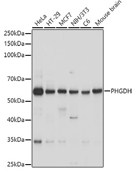
Western blot analysis of various lysates using PHGDH Rabbit pAb (CAB10461) at 1:1000 dilution. Secondary antibody: HRP-conjugated Goat anti-Rabbit IgG (H+L) (CABS014) at 1:10000 dilution. Lysates/proteins: 25μg per lane. Blocking buffer: 3% nonfat dry milk in TBST. Detection: ECL Basic Kit (AbGn00020). Exposure time: 1s.
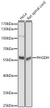
Western blot analysis of various lysates using PHGDH Rabbit pAb (CAB10461) at 1:1000 dilution. Secondary antibody: HRP-conjugated Goat anti-Rabbit IgG (H+L) (CABS014) at 1:10000 dilution. Lysates / proteins: 25 μg per lane. Blocking buffer: 3 % nonfat dry milk in TBST. Detection: ECL Basic Kit (AbGn00020). Exposure time: 5s.
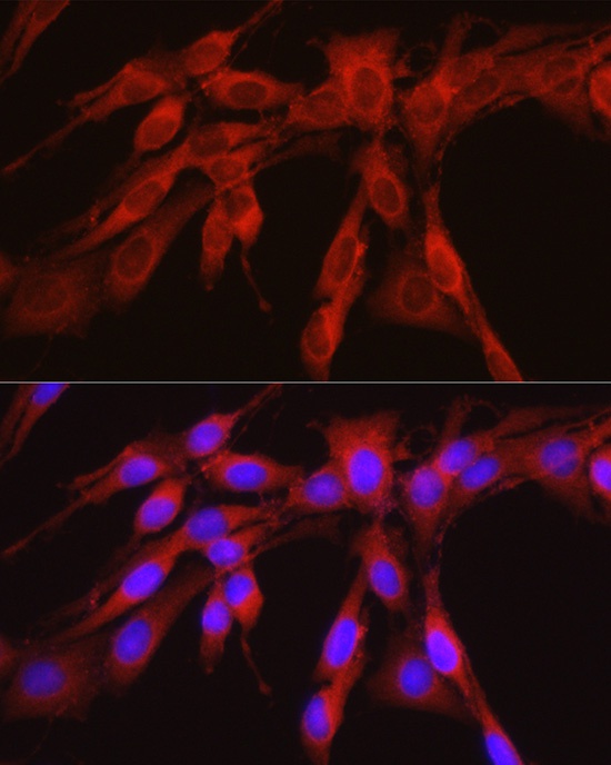
Immunofluorescence analysis of H9C2 cells using PHGDH Rabbit pAb (CAB10461) at dilution of 1:100. Blue: DAPI for nuclear staining.
Immunofluorescence analysis of U2OS cells using PHGDH Rabbit pAb (CAB10461) at dilution of 1:100. Blue: DAPI for nuclear staining.
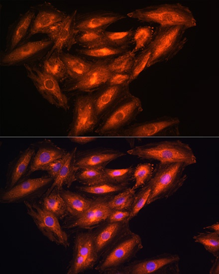
Immunofluorescence analysis of NIH/3T3 cells using PHGDH Rabbit pAb (CAB10461) at dilution of 1:300 (40x lens). Secondary antibody: Cy3-conjugated Goat anti-Rabbit IgG (H+L) (CABS007) at 1:500 dilution. Blue: DAPI for nuclear staining.
Immunofluorescence analysis of PC-12 cells using PHGDH Rabbit pAb (CAB10461) at dilution of 1:300 (40x lens). Secondary antibody: Cy3-conjugated Goat anti-Rabbit IgG (H+L) (CABS007) at 1:500 dilution. Blue: DAPI for nuclear staining.


")


