The Phospho-AMPKalpha1-T183+AMPKalpha2-T172 Monoclonal Antibody (CABP1441) is a high-quality antibody developed for reliable detection and analysis of target proteins. This antibody, developed using advanced monoclonal antibody technology, is reactive with both AMPK isoforms in various species, making it ideal for widespread use in experiments such as Western blotting and immunofluorescence.The phosphorylation of AMPK at threonine residues 183 and 172 is crucial for its activation and subsequent signaling pathways that regulate cellular energy balance, making this antibody particularly valuable for studies focusing on metabolic disorders and related conditions.
This antibody is validated for use in WB, ELISA applications and has demonstrated reactivity against Human, Mouse, Rat samples.
The protein encoded by this gene belongs to the ser/thr protein kinase family. It is the catalytic subunit of the 5'-prime-AMP-activated protein kinase (AMPK). AMPK is a cellular energy sensor conserved in all eukaryotic cells. The kinase activity of AMPK is activated by the stimuli that increase the cellular AMP/ATP ratio. AMPK regulates the activities of a number of key metabolic enzymes through phosphorylation. It protects cells from stresses that cause ATP depletion by switching off ATP-consuming biosynthetic pathways. Alternatively spliced transcript variants encoding distinct isoforms have been observed.
Purification Method
Affinity purification
Gene ID
5562 5563
Buffer Information
Store at -20℃. Avoid freeze / thaw cycles. Buffer: PBS with 0.09% Sodium azide,0.05% BSA,50% glycerol,pH7.3.
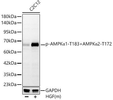
Western blot analysis of lysates from C2C12 cells, using Phospho-AMPKα1-T183+AMPKα2-T172 Rabbit mAb (CABP1441) at 1:1000 dilution. C2C12 cells were treated with mHGF(50ng/uL). Secondary antibody: HRP-conjugated Goat anti-Rabbit IgG (H+L) (CABS014) at 1:10000 dilution. Lysates/proteins: 25μg per lane. Blocking buffer: 3% nonfat dry milk in TBST. Detection: ECL Basic Kit (AbGn00020). Exposure time: 30s.
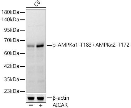
Western blot analysis of lysates from C6 cells, using Phospho-AMPKα1-T183+AMPKα2-T172 Rabbit mAb (CABP1441) at 1:1000 dilution. C6 cells were treated with AICAR (0. 5 mM) at 37℃ for 30 minutes after serum-starvation overnight. Secondary antibody: HRP-conjugated Goat anti-Rabbit IgG (H+L) (CABS014) at 1:10000 dilution. Lysates/proteins: 25μg per lane. Blocking buffer: 3% nonfat dry milk in TBST. Detection: ECL Basic Kit (AbGn00020). Exposure time: 30s.
")
")


