The Phospho-Histone H1.3-T17/Histone H1.4-T17 Monoclonal Antibody (CABP1132) is a high-quality antibody developed for reliable detection and analysis of target proteins. This antibody specifically recognizes phosphorylated histone H1.3 at threonine 17 and histone H1.4 at threonine 17, crucial modifications that play a role in chromatin structure and genome organization.Histone H1 is a key component of chromatin, acting as a linker histone that stabilizes nucleosome structure. The phosphorylation of histone H1 at specific sites, such as threonine 17 on H1.3 and H1.4, can impact transcriptional regulation and gene expression. By targeting these phosphorylation events, researchers can investigate the dynamic changes in chromatin structure that occur during processes like cell differentiation, development, and disease progression.
This antibody is validated for use in WB, ELISA applications and has demonstrated reactivity against Human, Mouse, Rat samples.
NIH/3T3 treated with Nocodazole, C6 treated with Nocodazole, HeLa treated with Nocodazole
Cellular Localization:
Nucleus.
Calculated MW:
30kDa
Observed MW:
30kDa
Histones are basic nuclear proteins responsible for nucleosome structure of the chromosomal fiber in eukaryotes. Two molecules of each of the four core histones (H2A, H2B, H3, and H4) form an octamer, around which approximately 146 bp of DNA is wrapped in repeating units, called nucleosomes. The linker histone, H1, interacts with linker DNA between nucleosomes and functions in the compaction of chromatin into higher order structures. This gene is intronless and encodes a replication-dependent histone that is a member of the histone H1 family. Transcripts from this gene lack polyA tails but instead contain a palindromic termination element. This gene is found in the large histone gene cluster on chromosome 6.
Purification Method
Affinity purification
Gene ID
3007 3008
RRID
AB_2864000
Buffer Information
Store at -20℃. Avoid freeze / thaw cycles. Buffer: PBS containing 50% glycerol and 0.05% BSA, preserved with proclin300 or sodium azide, pH 7.3.
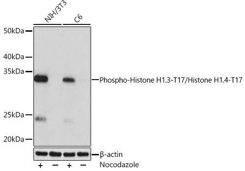
Western blot analysis of various lysates using Phospho-Histone H1.3-T17/Histone H1.4-T17 Rabbit mAb (CABP1132) at 1:1000 dilution. Both NIH/3T3 cells and C6 cells were treated with Nocodazole (50 ng/mL) at 37℃ for 20 hours. Secondary antibody: HRP-conjugated Goat anti-Rabbit IgG (H+L) (CABS014) at 1:10000 dilution. Lysates/proteins: 25 μg per lane. Blocking buffer: 3% BSA. Detection: ECL Basic Kit (AbGn00020). Exposure time: 1 s.
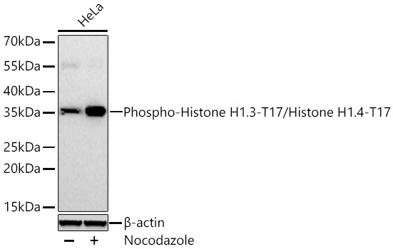
Western blot analysis of lysates from HeLa cells using Phospho-Histone H1.3-T17/Histone H1.4-T17 Rabbit mAb (CABP1132) at 1:2000 dilution incubated at room temperature for 1.5 hours. HeLa cells were treated with Nocodazole (100 ng/ml) at 37℃ for 16 hours. Secondary antibody: HRP-conjugated Goat anti-Rabbit IgG (H+L) (CABS014) at 1:10000 dilution. Lysates/proteins: 30 μg per lane. Blocking buffer: 3% nonfat dry milk in TBST. Detection: ECL Basic Kit (AbGn00020). Exposure time: 45 s.