The Phospho-p38 MAPK-T180/Y182 Monoclonal Antibody (CABP1311) is a high-quality antibody developed for reliable detection and analysis of target proteins. This gene encodes a member of a family of protein kinases that are involved in the integration of biochemical signals for a wide variety of cellular processes, including cell proliferation, differentiation, transcriptional regulation, and development. The encoded protein can be activated by proinflammatory cytokines and environmental stresses through phosphorylation by mitogen activated protein kinase kinases (MKKs). Alternative splicing results in multiple transcript variants. [provided by RefSeq, Mar 2014]
This antibody is validated for use in WB, ELISA applications and has demonstrated reactivity against Human, Mouse, Rat samples.
Product Name:
Phospho-p38 MAPK-T180/Y182 Monoclonal Antibody
SKU:
CABP1311
Size:
100μL, 20μL
Reactivity:
Human, Mouse, Rat
Clone Number:
ARC51018
Immunogen:
Synthetic peptide. This information is considered to be commercially sensitive.
Tested Applications:
WBELISA
Recommended Dilution:
WB
1:2000 - 1:20000
ELISA
Recommended starting concentration is 1 μg/mL. Please optimize the concentration based on your specific assay requirements.
293T treated with UV, NIH/3T3 treated with UV, C6 treated with UV
Cellular Localization:
Cytoplasm, Cytosol, Nucleoplasm, Nucleus.
Calculated MW:
29kDa/34kDa/35kDa/41kDa
Observed MW:
43kDa
This gene encodes a member of a family of protein kinases that are involved in the integration of biochemical signals for a wide variety of cellular processes, including cell proliferation, differentiation, transcriptional regulation, and development. The encoded protein can be activated by proinflammatory cytokines and environmental stresses through phosphorylation by mitogen activated protein kinase kinases (MKKs). Alternative splicing results in multiple transcript variants. [provided by RefSeq, Mar 2014]
Purification Method
Affinity purification
Gene ID
5600 6300 5603 1432
Buffer Information
Store at -20℃. Avoid freeze / thaw cycles. Buffer: PBS containing 50% glycerol and 0.05% BSA, preserved with proclin300 or sodium azide, pH 7.3.
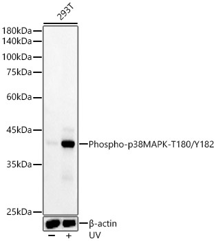
Western blot analysis of various lysates, using Phospho-p38 MAPK-T180/Y182 Rabbit mAb (CABP1311) at 1:20000 dilution. 293T cells were treated with UV at room temperature for 15-30 minutes. Secondary antibody: HRP-conjugated Goat anti-Rabbit IgG (H+L) (AS014) at 1:10000 dilution. Lysates/proteins: 25μg per lane. Blocking buffer: 3% nonfat dry milk in TBST. Detection: ECL Basic Kit (AbGn00020). Exposure time: 10s.
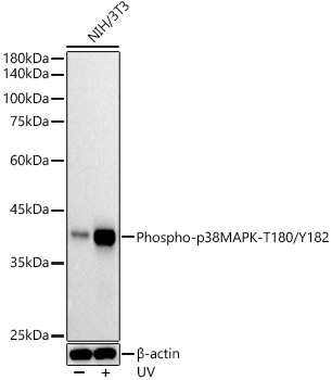
Western blot analysis of lysates from NIH/3T3 cells, using Phospho-p38 MAPK-T180/Y182 Rabbit mAb (CABP1311) at 1:20000 dilution. NIH/3T3 cells were treated with UV at room temperature for 15-30 minutes. Secondary antibody: HRP-conjugated Goat anti-Rabbit IgG (H+L) (AS014) at 1:10000 dilution. Lysates/proteins: 25μg per lane. Blocking buffer: 3% nonfat dry milk in TBST. Detection: ECL Basic Kit (AbGn00020). Exposure time: 10s.
 at 1:20000 dilution. 293T cells were treated by UV at room temperature for 15-30 minutes. Secondary antibody: HRP Goat Anti-Rabbit IgG (H+L) at 1:10000 dilution. Lysates/proteins: 25μg per lane. Blocking buffer: 3% nonfat dry milk in TBST.")
 at 1:20000 dilution. 293T cells were treated by UV at room temperature for 15-30 minutes. Secondary antibody: HRP Goat Anti-Rabbit IgG (H+L) at 1:10000 dilution. Lysates/proteins: 25μg per lane. Blocking buffer: 3% nonfat dry milk in TBST.")

")

