The Phospho-PDPK1-S241 Antibody (CABP0477) is a high-quality antibody developed for reliable detection and analysis of target proteins. The antibody, raised in rabbits, is highly specific and reactive towards human samples, making it ideal for Western blot applications. It targets the phosphorylated form of PDK1 at serine 241, allowing for precise detection and analysis in various cell types.Phosphorylation of PDK1 at serine 241 plays a crucial role in the activation of downstream signaling cascades involved in cell growth, proliferation, and metabolism.
This antibody is validated for use in WB, IF/ICC, ELISA applications and has demonstrated reactivity against Human, Mouse, Rat samples.
Product Name:
Phospho-PDPK1-S241 Antibody
SKU:
CABP0477
Size:
20μL, 100μL
Reactivity:
Human, Mouse, Rat
Conjugate:
Unconjugated
Immunogen:
Synthetic peptide. This information is considered to be commercially sensitive.
Sequence:
ANSF V
Tested Applications:
WBIF/ICCELISA
Recommended Dilution:
WB
1:500 - 1:2000
IF/ICC
1:50 - 1:200
ELISA
Recommended starting concentration is 1 μg/mL. Please optimize the concentration based on your specific assay requirements.
Enables 3-phosphoinositide-dependent protein kinase activity; phospholipase activator activity; and phospholipase binding activity. Involved in several processes, including cell surface receptor signaling pathway; regulation of protein kinase activity; and regulation of signal transduction. Acts upstream of or within intracellular signal transduction. Located in cell projection; cytosol; and plasma membrane. Implicated in prostate cancer. Biomarker of lung non-small cell carcinoma.
Purification Method
Affinity purification
Gene ID
5170
RRID
AB_2771409
Buffer Information
Store at -20℃. Avoid freeze / thaw cycles. Buffer: PBS with 0.09% Sodium azide,50% glycerol,pH7.3.
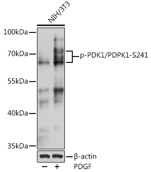
Western blot analysis of lysates from NIH/3T3 cells, using Phospho-PDK1/PDPK1-S241 pAb (CABP0477) at 1:1000 dilution. NIH/3T3 cells were treated with PDGF (100 ng/mL) at 37℃ for 30 minutes after serum-starvation overnight. Secondary antibody: HRP-conjugated Goat anti-Rabbit IgG (H+L) (CABS014) at 1:10000 dilution. Lysates/proteins: 25μg per lane. Blocking buffer: 3% BSA. Detection: ECL Basic Kit (AbGn00020). Exposure time: 60s.
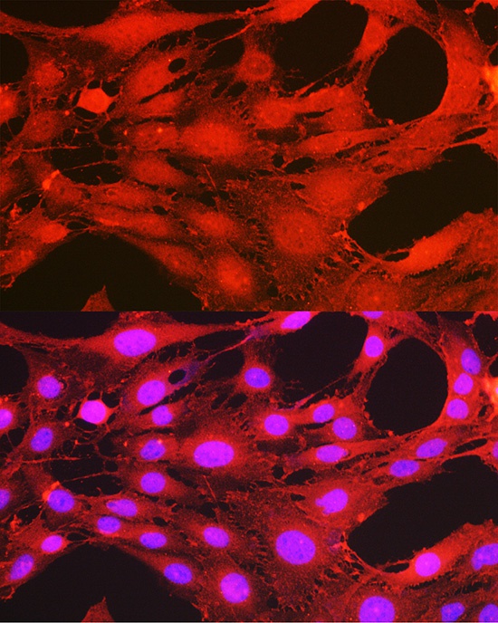
Immunofluorescence analysis of C6 cells using Phospho-PDK1/PDPK1-S241 Rabbit pAb (CABP0477) at dilution of 1:100 (40x lens). Secondary antibody: Cy3-conjugated Goat anti-Rabbit IgG (H+L) (CABS007) at 1:500 dilution. Blue: DAPI for nuclear staining.
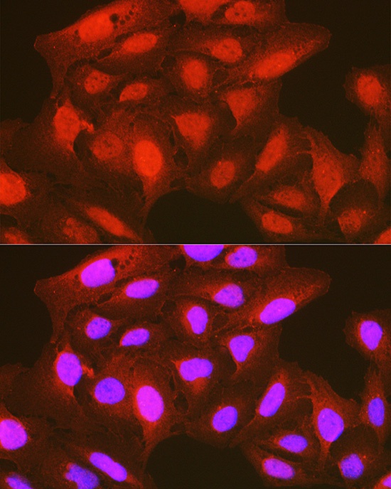
Immunofluorescence analysis of U-2 OS cells using Phospho-PDK1/PDPK1-S241 Rabbit pAb (CABP0477) at dilution of 1:100 (40x lens). Secondary antibody: Cy3-conjugated Goat anti-Rabbit IgG (H+L) (CABS007) at 1:500 dilution. Blue: DAPI for nuclear staining.