The PLSCR3 Antibody (CAB12371) is a high-quality antibody developed for reliable detection and analysis of target proteins. PLSCR3 is involved in phospholipid translocation and cell membrane dynamics, playing a crucial role in various cellular processes such as apoptosis, cell signaling, and lipid metabolism.This antibody, produced in rabbits, is highly specific and reactive with human samples, making it suitable for use in a variety of applications including Western blotting, immunofluorescence, and immunohistochemistry. By binding to the PLSCR3 protein, this antibody enables researchers to detect and analyze the expression levels and localization of PLSCR3 in different cell types and tissues.
This antibody is validated for use in WB, IF/ICC, ELISA applications and has demonstrated reactivity against Human, Mouse, Rat samples.
Product Name:
PLSCR3 Antibody
SKU:
CAB12371
Size:
20μL, 100μL
Reactivity:
Human, Mouse, Rat
Conjugate:
Unconjugated
Immunogen:
Recombinant protein (or fragment).This information is considered to be commercially sensitive.
Mitochondrion Membrane, Single-Pass Type Ii Membrane Protein.
Calculated MW:
32kDa
Observed MW:
32kDa
Enables several functions, including calcium-dependent protein binding activity; metal ion binding activity; and phospholipid scramblase activity. Involved in several processes, including cardiolipin biosynthetic process; regulation of apoptotic process; and regulation of release of cytochrome c from mitochondria. Located in cytosol; mitochondrion; and plasma membrane.
Purification Method
Affinity purification
Gene ID
57048
RRID
AB_2759215
Buffer Information
Store at -20℃. Avoid freeze / thaw cycles. Buffer: PBS with 0.01% thimerosal,50% glycerol,pH7.3.
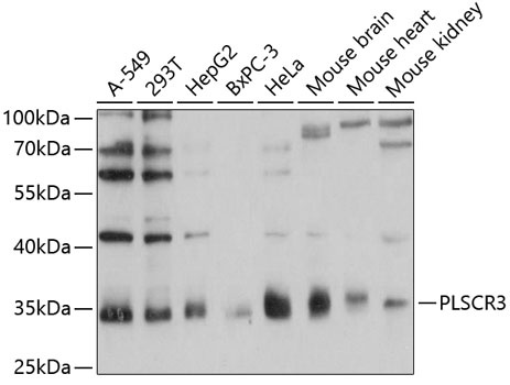
Western blot analysis of various lysates using PLSCR3 Rabbit pAb (CAB12371) at 1:1000 dilution. Secondary antibody: HRP-conjugated Goat anti-Rabbit IgG (H+L) (CABS014) at 1:10000 dilution. Lysates/proteins: 25μg per lane. Blocking buffer: 3% nonfat dry milk in TBST. Detection: ECL Basic Kit (AbGn00020). Exposure time: 1s.
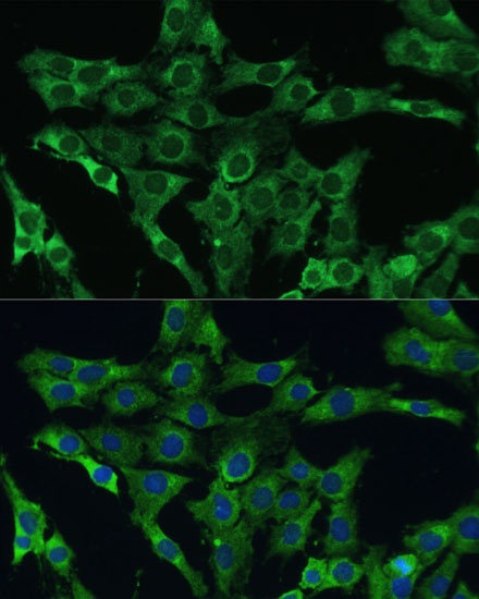
Immunofluorescence analysis of C6 cells using PLSCR3 Rabbit pAb (CAB12371) at dilution of 1:100 (40x lens). Secondary antibody: Cy3-conjugated Goat anti-Rabbit IgG (H+L) (CABS007) at 1:500 dilution. Blue: DAPI for nuclear staining.


 at 1:10000 dilution. Lysates/proteins: 25ug per lane. Blocking buffer: 3% nonfat dry milk in TBST. Detection: ECL Basic Kit. Exposure time: 180s.")
 at 1:10000 dilution. Lysates/proteins: 25ug per lane. Blocking buffer: 3% nonfat dry milk in TBST. Detection: ECL Basic Kit. Exposure time: 30s.")


