The PNKP Antibody (CAB6693) is a high-quality antibody developed for reliable detection and analysis of target proteins. This antibody, generated in rabbits, exhibits high reactivity with human samples and has been validated for use in Western blot applications. By binding specifically to the PNKP protein, this antibody enables accurate detection and analysis in a variety of cell types, making it an ideal choice for investigations in molecular biology and genetics.
This antibody is validated for use in WB, IHC-P, IF/ICC, ELISA applications and has demonstrated reactivity against Human, Mouse, Rat samples.
Product Name:
PNKP Antibody
SKU:
CAB6693
Size:
20μL, 100μL
Reactivity:
Human, Mouse, Rat
Conjugate:
Unconjugated
Immunogen:
Recombinant protein (or fragment).This information is considered to be commercially sensitive.
Recommended starting concentration is 1 μg/mL. Please optimize the concentration based on your specific assay requirements.
Synonyms:
PNK, AOA4, MCSZ, CMT2B2, EIEE10, PNKP
Positive Sample:
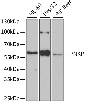
HL-60, HepG2, Rat liver
Cellular Localization:
Nucleus.
Calculated MW:
57kDa
Observed MW:
57kDa
This locus represents a gene involved in DNA repair. In response to ionizing radiation or oxidative damage, the protein encoded by this locus catalyzes 5' phosphorylation and 3' dephosphorylation of nucleic acids. Mutations at this locus have been associated with microcephaly, seizures, and developmental delay.
Purification Method
Affinity purification
Gene ID
11284
RRID
AB_2767277
Buffer Information
Store at -20℃. Avoid freeze / thaw cycles. Buffer: PBS containing 50% glycerol, preserved with proclin300 or sodium azide, pH 7.3.
Western blot analysis of various lysates using PNKP Rabbit pAb (CAB6693) at 1:1000 dilution. Secondary antibody: HRP-conjugated Goat anti-Rabbit IgG (H+L) (CABS014) at 1:10000 dilution. Lysates/proteins: 25μg per lane. Blocking buffer: 3% nonfat dry milk in TBST. Detection: ECL Basic Kit (AbGn00020). Exposure time: 90s.
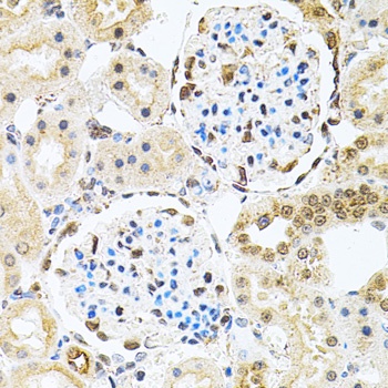
Immunohistochemistry analysis of paraffin-embedded Rat kidney using PNKP Rabbit pAb (CAB6693) at dilution of 1:100 (40x lens). Microwave antigen retrieval performed with 0.01M PBS Buffer (pH 7.2) prior to IHC staining.
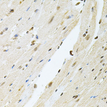
Immunohistochemistry analysis of paraffin-embedded Mouse heart using PNKP Rabbit pAb (CAB6693) at dilution of 1:100 (40x lens). Microwave antigen retrieval performed with 0.01M PBS Buffer (pH 7.2) prior to IHC staining.
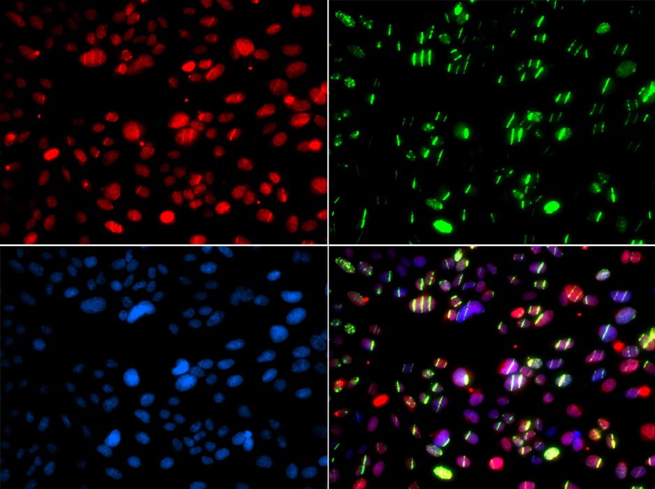
Immunofluorescence analysis of GFP-RNF168 transgenic U2OS cells using PNKP Rabbit pAb (CAB6693). Green:GFP-RNF168 fusion protein expression for DNA damage marker.Secondary antibody: Cy3-conjugated Goat anti-Rabbit IgG (H+L) (CABS007) at 1:500 dilution. Blue: DAPI for nuclear staining. RNF168(GFP) can be used to mark cells damaged by UV-A laser for they always gather around DNA damage region.