The PRKCQ Antibody (CAB5834) is a high-quality antibody developed for reliable detection and analysis of target proteins. This antibody, produced in rabbits, has been extensively validated for Western blot applications and is highly reactive with human samples. By specifically binding to PRKCQ, this antibody allows for the detection and analysis of PRKCQ in various cell types, making it ideal for studies in immunology and cancer research.
This antibody is validated for use in WB, ELISA applications and has demonstrated reactivity against Human, Mouse, Rat samples.
Product Name:
PRKCQ Antibody
SKU:
CAB5834
Size:
20μL, 100μL
Reactivity:
Human, Mouse, Rat
Conjugate:
Unconjugated
Immunogen:
Recombinant protein (or fragment).This information is considered to be commercially sensitive.
Protein kinase C (PKC) is a family of serine- and threonine-specific protein kinases that can be activated by calcium and the second messenger diacylglycerol. PKC family members phosphorylate a wide variety of protein targets and are known to be involved in diverse cellular signaling pathways. PKC family members also serve as major receptors for phorbol esters, a class of tumor promoters. Each member of the PKC family has a specific expression profile and is believed to play a distinct role. The protein encoded by this gene is one of the PKC family members. It is a calcium-independent and phospholipid-dependent protein kinase. This kinase is important for T-cell activation. It is required for the activation of the transcription factors NF-kappaB and AP-1, and may link the T cell receptor (TCR) signaling complex to the activation of the transcription factors.
Purification Method
Affinity purification
Gene ID
5588
RRID
AB_2766584
Buffer Information
Store at -20℃. Avoid freeze / thaw cycles. Buffer: PBS containing 50% glycerol, preserved with proclin300 or sodium azide, pH 7.3.
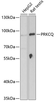
Western blot analysis of various lysates using PRKCQ Rabbit pAb (CAB5834) at 1:1000 dilution. Secondary antibody: HRP-conjugated Goat anti-Rabbit IgG (H+L) (CABS014) at 1:10000 dilution. Lysates/proteins: 25μg per lane. Blocking buffer: 3% nonfat dry milk in TBST. Detection: ECL Basic Kit (AbGn00020). Exposure time: 90s.