The JTB Antibody (CAB10427) is a high-quality antibody developed for reliable detection and analysis of target proteins. This antibody, generated in rabbits, has high reactivity with human samples and is validated for use in Western blot applications. By binding to the JTB protein, this antibody enables researchers to detect and analyze JTB expression in various cell types, making it an essential tool for studies in epigenetics and cancer research.
This antibody is validated for use in WB, ELISA applications and has demonstrated reactivity against Human, Rat samples.
Product Name:
JTB Antibody
SKU:
CAB10427
Size:
20μL, 100μL
Reactivity:
Human, Rat
Conjugate:
Unconjugated
Immunogen:
Recombinant protein (or fragment).This information is considered to be commercially sensitive.
Recommended starting concentration is 1 μg/mL. Please optimize the concentration based on your specific assay requirements.
Synonyms:
PAR, hJT, HJTB, HSPC222, JTB
Positive Sample:
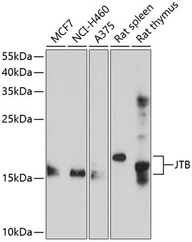
MCF7, NCI-H460, A375, Rat spleen, Rat thymus
Cellular Localization:
Cytoplasm, Membrane, Mitochondrion, Single-Pass Type I Membrane Protein, Centrosome, Cytoskeleton, Microtubule Organizing Center, Spindle.
Calculated MW:
16kDa
Observed MW:
16kDa
Enables protein kinase binding activity. Involved in mitotic cytokinesis and positive regulation of protein kinase activity. Located in cytoplasm and midbody. Colocalizes with centrosome and spindle.
Purification Method
Affinity purification
Gene ID
10899
RRID
AB_2757974
Buffer Information
Store at -20℃. Avoid freeze / thaw cycles. Buffer: PBS containing 50% glycerol, preserved with proclin300 or sodium azide, pH 7.3.
Western blot analysis of various lysates using JTB Rabbit pAb (CAB10427) at 1:1000 dilution. Secondary antibody: HRP-conjugated Goat anti-Rabbit IgG (H+L) (CABS014) at 1:10000 dilution. Lysates/proteins: 25μg per lane. Blocking buffer: 3% nonfat dry milk in TBST. Detection: ECL Enhanced Kit (AbGn00021). Exposure time: 60s.