The PRPF8 Monoclonal Antibody (CAB4575) is a high-quality antibody developed for reliable detection and analysis of target proteins. This antibody, produced in rabbits, is highly specific and reactive with human samples, making it ideal for use in Western blot applications. By binding to PRPF8, this antibody enables precise detection and analysis of the protein in various cell types, facilitating research in fields such as molecular biology, genetics, and RNA splicing mechanisms.
This antibody is validated for use in WB, IHC-P, IF/ICC, ELISA applications and has demonstrated reactivity against Human, Mouse, Rat samples.
Product Name:
PRPF8 Monoclonal Antibody
SKU:
CAB4575
Size:
20μL, 100μL
Reactivity:
Human, Mouse, Rat
Clone Number:
ARC1038
Conjugate:
Unconjugated
Immunogen:
Recombinant protein (or fragment).This information is considered to be commercially sensitive.
Recommended starting concentration is 1 μg/mL. Please optimize the concentration based on your specific assay requirements.
Synonyms:
PRP8, RP13, HPRP8, PRPC8, SNRNP220, PRPF8
Positive Sample:
HeLa, 293T, Jurkat, Mouse brain, Mouse spleen
Cellular Localization:
Nucleus Speckle.
Calculated MW:
274kDa
Observed MW:
274kDa
Pre-mRNA splicing occurs in 2 sequential transesterification steps. The protein encoded by this gene is a component of both U2- and U12-dependent spliceosomes, and found to be essential for the catalytic step II in pre-mRNA splicing process. It contains several WD repeats, which function in protein-protein interactions. This protein has a sequence similarity to yeast Prp8 protein. This gene is a candidate gene for autosomal dominant retinitis pigmentosa.
Purification Method
Affinity purification
Gene ID
10594
RRID
AB_2863300
Buffer Information
Store at -20℃. Avoid freeze / thaw cycles. Buffer: PBS containing 50% glycerol and 0.05% BSA, preserved with proclin300 or sodium azide, pH 7.3.
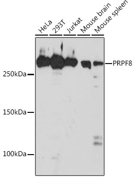
Western blot analysis of various lysates using PRPF8 Rabbit mAb (CAB4575) at 1:1000 dilution. Secondary antibody: HRP-conjugated Goat anti-Rabbit IgG (H+L) (CABS014) at 1:10000 dilution. Lysates/proteins: 25μg per lane. Blocking buffer: 3% nonfat dry milk in TBST. Detection: ECL Basic Kit (AbGn00020). Exposure time: 30s.
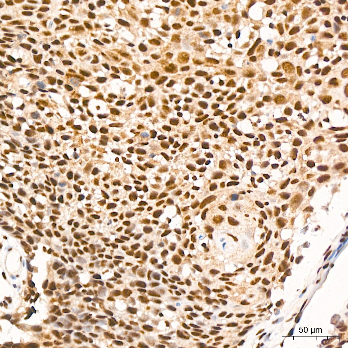
Immunohistochemistry analysis of paraffin-embedded Human cervix cancer tissue using PRPF8 Rabbit mAb (CAB4575) at a dilution of 1:200 (40x lens). High pressure antigen retrieval was performed with 0.01 M citrate buffer (pH 6.0) prior to IHC staining.
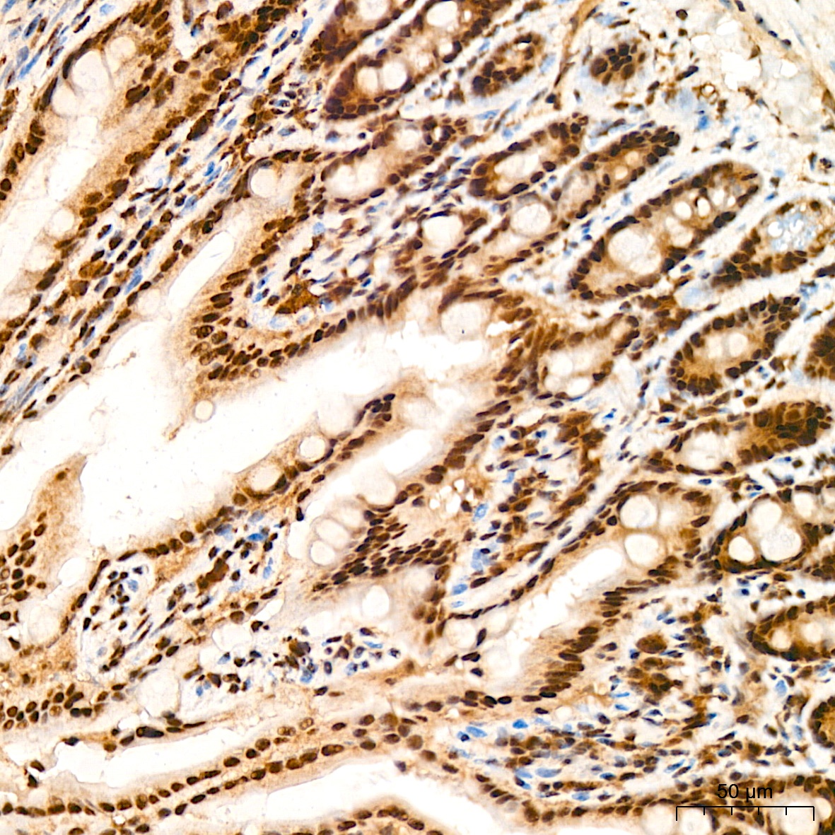
Immunohistochemistry analysis of paraffin-embedded Mouse colon tissue using PRPF8 Rabbit mAb (CAB4575) at a dilution of 1:200 (40x lens). High pressure antigen retrieval was performed with 0.01 M citrate buffer (pH 6.0) prior to IHC staining.
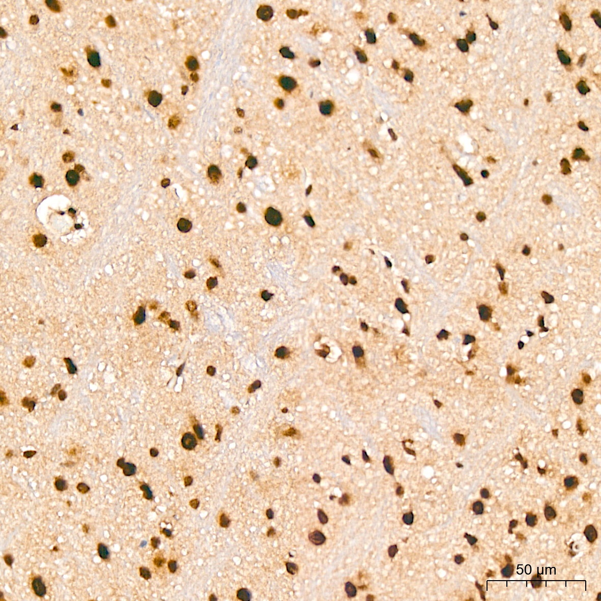
Immunohistochemistry analysis of paraffin-embedded Mouse brain tissue using PRPF8 Rabbit mAb (CAB4575) at a dilution of 1:200 (40x lens). High pressure antigen retrieval was performed with 0.01 M citrate buffer (pH 6.0) prior to IHC staining.
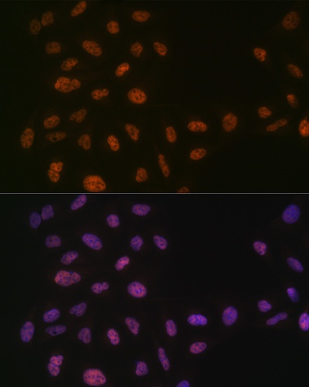
Immunofluorescence analysis of U-2 OS cells using PRPF8 Rabbit mAb (CAB4575) at dilution of 1:100 (40x lens). Secondary antibody: Cy3-conjugated Goat anti-Rabbit IgG (H+L) (CABS007) at 1:500 dilution. Blue: DAPI for nuclear staining.