The [KO Validated] SHP2 Antibody (CAB12486) is a high-quality antibody developed for reliable detection and analysis of target proteins. This antibody, produced in rabbits, targets PTPN11 and is validated for use in Western blot applications with human samples. By specifically binding to PTPN11, researchers can detect and analyze this critical protein in different cell types, making it suitable for studies in cell signaling, cancer biology, and developmental biology.
This antibody is validated for use in WB, IF/ICC, IP, ELISA applications and has demonstrated reactivity against Human, Mouse, Rat samples.
Product Name:
[KO Validated] SHP2 Antibody
SKU:
CAB12486
Size:
20μL, 100μL
Reactivity:
Human, Mouse, Rat
Conjugate:
Unconjugated
Immunogen:
Recombinant protein (or fragment).This information is considered to be commercially sensitive.
U-87MG, Jurkat, A-431, MCF-7, Mouse brain, Rat brain, Rat lung
Cellular Localization:
Cytoplasm.
Calculated MW:
68kDa
Observed MW:
75kDa
The protein encoded by this gene is a member of the protein tyrosine phosphatase (PTP) family. PTPs are known to be signaling molecules that regulate a variety of cellular processes including cell growth, differentiation, mitotic cycle, and oncogenic transformation. This PTP contains two tandem Src homology-2 domains, which function as phospho-tyrosine binding domains and mediate the interaction of this PTP with its substrates. This PTP is widely expressed in most tissues and plays a regulatory role in various cell signaling events that are important for a diversity of cell functions, such as mitogenic activation, metabolic control, transcription regulation, and cell migration. Mutations in this gene are a cause of Noonan syndrome as well as acute myeloid leukemia.
Purification Method
Affinity purification
Gene ID
5781
RRID
AB_2861667
Buffer Information
Store at -20℃. Avoid freeze / thaw cycles. Buffer: PBS containing 50% glycerol, preserved with proclin300 or sodium azide, pH 7.3.
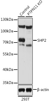
Western blot analysis of lysates from wild type (WT) and SHP2 knockout (KO) 293T cells, using [KO Validated] SHP2 Rabbit pAb (CAB12486) at 1:1000 dilution. Secondary antibody: HRP-conjugated Goat anti-Rabbit IgG (H+L) (CABS014) at 1:10000 dilution. Lysates/proteins: 25μg per lane. Blocking buffer: 3% nonfat dry milk in TBST. Detection: ECL Basic Kit (AbGn00020). Exposure time: 30s.
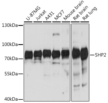
Western blot analysis of various lysates using [KO Validated] SHP2 Rabbit pAb (CAB12486) at 1:1000 dilution. Secondary antibody: HRP-conjugated Goat anti-Rabbit IgG (H+L) (CABS014) at 1:10000 dilution. Lysates/proteins: 25μg per lane. Blocking buffer: 3% nonfat dry milk in TBST. Detection: ECL Basic Kit (AbGn00020). Exposure time: 3s.
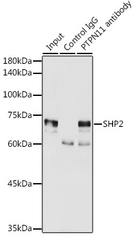
Immunoprecipitation analysis of 300 μg extracts of MCF7 cells using 3 μg SHP2 antibody (CAB12486). Western blot was performed from the immunoprecipitate using SHP2 antibody (CAB12486) at a dilution of 1:2000.