The RAB7 Monoclonal Antibody (CAB12308) is a high-quality antibody developed for reliable detection and analysis of target proteins. This antibody, generated using rabbit monoclonal technology, is highly specific and reactive with human samples, making it an essential tool for studying Rab7 in various cellular processes.Rab7 is known to be involved in the late endocytic pathway, lysosome biogenesis, and autophagy, making it a crucial player in cellular homeostasis and signaling. By targeting Rab7 with this antibody, researchers can effectively detect and analyze its expression and localization in different cell types, providing valuable insights into its function and potential therapeutic targets.
This antibody is validated for use in WB, IHC-P, IF/ICC, IP, ELISA applications and has demonstrated reactivity against Human, Mouse, Rat samples.
Product Name:
RAB7 Monoclonal Antibody
SKU:
CAB12308
Size:
20μL, 100μL
Reactivity:
Human, Mouse, Rat
Clone Number:
ARC54437
Conjugate:
Unconjugated
Immunogen:
Synthetic peptide. This information is considered to be commercially sensitive.
RAB family members are small, RAS-related GTP-binding proteins that are important regulators of vesicular transport. Each RAB protein targets multiple proteins that act in exocytic / endocytic pathways. This gene encodes a RAB family member that regulates vesicle traffic in the late endosomes and also from late endosomes to lysosomes. This encoded protein is also involved in the cellular vacuolation of the VacA cytotoxin of Helicobacter pylori. Mutations at highly conserved amino acid residues in this gene have caused some forms of Charcot-Marie-Tooth (CMT) type 2 neuropathies.
Purification Method
Affinity purification
Gene ID
7879
RRID
AB_2861652
Buffer Information
Store at -20℃. Avoid freeze / thaw cycles. Buffer: PBS containing 50% glycerol and 0.05% BSA, preserved with proclin300 or sodium azide, pH 7.3.
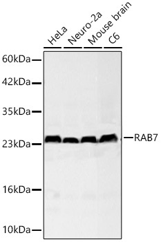
Western blot analysis of various lysates using RAB7 Rabbit mAb (CAB12308) at 1:55000 dilution incubated overnight at 4℃. Secondary antibody: HRP-conjugated Goat anti-Rabbit IgG (H+L) (CABS014) at 1:10000 dilution. Lysates/proteins: 25 μg per lane. Blocking buffer: 3% nonfat dry milk in TBST. Detection: ECL Basic Kit (AbGn00020). Exposure time: 20s.
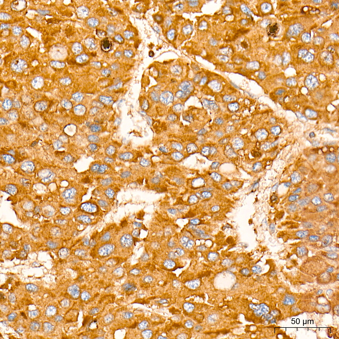
Immunohistochemistry analysis of paraffin-embedded Human liver cancer tissue using RAB7 Rabbit mAb (CAB12308) at a dilution of 1:3000 (40x lens). High pressure antigen retrieval performed with 0.01M Tris-EDTA Buffer (pH 9.0) prior to IHC staining.
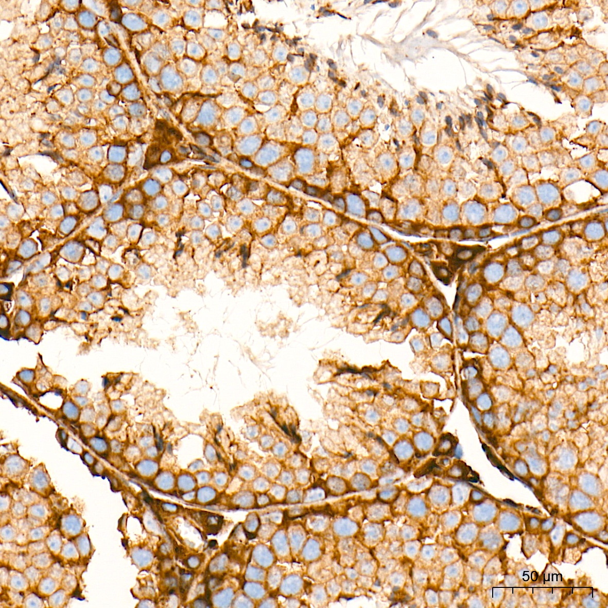
Immunohistochemistry analysis of paraffin-embedded Mouse testis tissue using RAB7 Rabbit mAb (CAB12308) at a dilution of 1:3000 (40x lens). High pressure antigen retrieval performed with 0.01M Tris-EDTA Buffer (pH 9.0) prior to IHC staining.
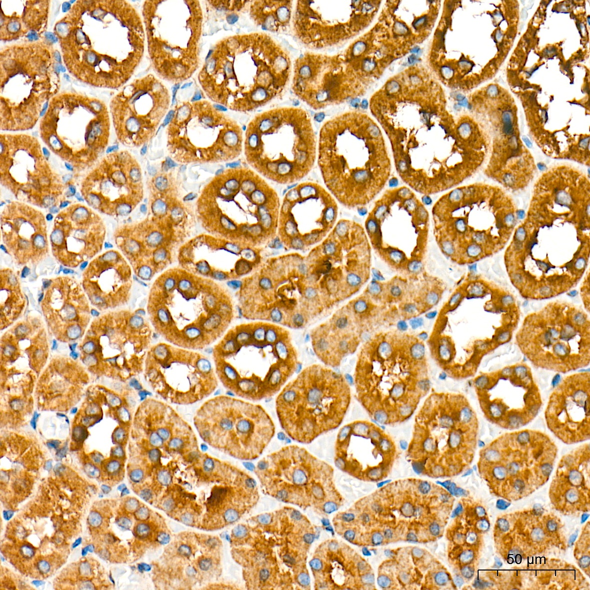
Immunohistochemistry analysis of paraffin-embedded Rat kidney tissue using RAB7 Rabbit mAb (CAB12308) at a dilution of 1:3000 (40x lens). High pressure antigen retrieval performed with 0.01M Tris-EDTA Buffer (pH 9.0) prior to IHC staining.
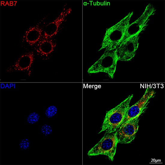
Confocal imaging of NIH/3T3 cells using RAB7 Rabbit mAb (CAB12308, dilution 1:2000) followed by a further incubation with Cy3 Goat Anti-Rabbit IgG (H+L) (CABS007, dilution 1:500) (Red). The cells were counterstained with α-Tubulin Mouse mAb (AC012, dilution 1:400) followed by incubation with ABflo® 488-conjugated Goat Anti-Mouse IgG (H+L) Ab (CABS076, dilution 1:500) (Green). DAPI was used for nuclear staining (Blue). Objective: 100x.
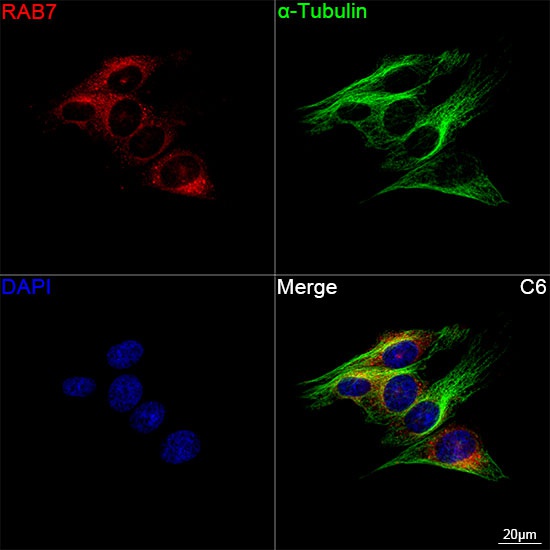
Confocal imaging of C6 cells using RAB7 Rabbit mAb (CAB12308, dilution 1:2000) followed by a further incubation with Cy3 Goat Anti-Rabbit IgG (H+L) (CABS007, dilution 1:500) (Red). The cells were counterstained with α-Tubulin Mouse mAb (AC012, dilution 1:400) followed by incubation with ABflo® 488-conjugated Goat Anti-Mouse IgG (H+L) Ab (CABS076, dilution 1:500) (Green). DAPI was used for nuclear staining (Blue). Objective: 100x.